. 2003 Jun;162(6):1817-22.
doi: 10.1016/S0002-9440(10)64316-7.

Protease-activated receptor-2 signaling triggers dendritic cell development

Affiliations

Protease-activated receptor-2 signaling triggers dendritic cell development

Ryan C Fields et al. Am J Pathol. 2003 Jun.

Abstract

Dendritic cells (DC) are potent antigen-presenting cells that govern the effector cell responses of the immune system. DC are thought to continuously develop from circulating progenitors in a process that is accelerated by inflammatory stimuli. However, the physiological signals that regulate the development of DC from precursor cells have not been well defined. Here we show that a serine protease acting via protease-activated receptor-2 (PAR-2) stimulates the development of DC from bone marrow progenitor cells cultured in granulocyte-macrophage colony-stimulating factor and IL-4. DC fail to develop in bone marrow cultures treated with soy bean trypsin inhibitor, a serine protease inhibitor, but this inhibition is overcome by a PAR-2 agonist peptide. DC do not spontaneously develop from the bone marrow of PAR-2-deficient mice, but can be stimulated to do so by inflammatory mediators. These results suggest that endogenous serine proteases stimulate DC development in vitro. Thus, serine proteases may help trigger adaptive immune responses in vivo.


Figures

Figure 1.
Figure 1.
Inhibition of serine proteases in DC media blocks DC development. Cells cultured in GM-CSF and IL-4 demonstrate high levels of DC surface markers (a) and show multiple cytoplasmic extensions and abundant cytoplasm (b, c). Cells cultured in GM-CSF and IL-4 supplemented with SBTI demonstrate low levels of DC markers (d), large nuclei, and multiple vacuoles (e, f). g: DC surface markers (▪ = I-Ab, ▴ = CD11c, ▾ = CD80, ♦ = CD86) decrease dose-dependently when SBTI is added to cultures. Results are shown as percent-positive expression by flow cytometry based on 20,000 cell counts. h: Cells not treated with SBTI (▪) demonstrate an increased ability to stimulate a mixed leukocyte reaction when compared to SBTI treated cells (▪). Data were significant (P < 0.03) in all stimulator-to-responder ratios. i: SBTI treated cells (▪) showed an increased ability to take up FITC-labeled dextran when compared to non-SBTI treated cells (▴). Scale bars, 15 μm; b and e and c and f are equivalent in magnification.
Figure 2.
Figure 2.
Stimulation of SBTI-treated bone marrow progenitor cells triggers DC development. When either TNF-α (a) or a crosslinking CD40 antibody (b) are added to SBTI treated cultures 24 hours before harvest, high levels of all surface markers and DC morphology are observed. Micron bar, 15 μm; all micrographs are equivalent in magnification.
Figure 3.
Figure 3.
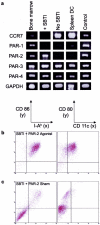
Stimulation of the PAR-2 receptor on SBTI-treated bone marrow progenitor cells triggers DC development. a: The PAR-2 receptor is expressed on SBTI-treated DC, but not on CCR7 high DC cultured from bone marrow and harvested from spleen (and further stimulated with TNF-α). RT-PCR expression of CCR7, PAR-1, -2, -3, -4, and GAPDH in SBTI- and non-SBTI-treated day 4 DC, spleen DC, fresh bone marrow progenitor cells, and control tissues (spleen for CCR7 and PAR-2, heart muscle for PAR-1, -3, and -4, and GAPDH). Results were similar across all cycle reactions. b and c: SBTI-treated cells display DC surface phenotype and morphology in response to PAR-2 stimulation. SBTI treated cells were treated with 10 μmol/L PAR-2 agonist (b) or PAR-2 agonist control (c) peptide 24 hours before harvest.
Figure 4.
Figure 4.
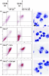
Generation of DC from bone marrow progenitor cells of PAR-2−/− mice is impaired. a: Culturing bone marrow progenitor cells from PAR-2+/+ mice in GM-CSF and IL-4 results in mature DC surface phenotype and morphology. b: Culturing bone marrow progenitor cells from PAR-2−/− mice does not result in mature DC surface phenotype or morphology. c and d: The addition of TNF-α (c) or a crosslinking CD40 antibody (d) to PAR-2−/− cultures 24 hours before harvest triggers DC development as indicated by surface phenotype and morphology. Micron bar, 15 μm; all micrographs are equivalent in magnification.

Similar articles

Cited by

References

    1. Steinman RM: The dendritic cell system and its role in immunogenicity. Annu Rev Immunol 1991, 9:271-296 - PubMed
    1. Banchereau J, Steinman RM: Dendritic cells and the control of immunity. Nature 1998, 392:245-252 - PubMed
    1. Cella M, Engering A, Pinet V, Pieters J, Lanzavecchia A: Inflammatory stimuli induce accumulation of MHC class II complexes on dendritic cells. Nature 1997, 388:782-787 - PubMed
    1. McWilliam AS, Napoli S, Marsh AM, Pemper FL, Nelson DJ, Pimm CL, Stumbles PA, Wells TN, Holt PG: Dendritic cells are recruited into the airway epithelium during the inflammatory response to a broad spectrum of stimuli. J Exp Med 1996, 184:2429-2432 - PMC - PubMed
    1. Matzinger P: An innate sense of danger. Semin Immunol 1998, 10:399-415 - PubMed

Publication types

MeSH terms

Substances

LinkOut - more resources