. 2003 Jun;162(6):1831-43.
doi: 10.1016/S0002-9440(10)64318-0.

Matrilysin (matrix metalloproteinase-7) mediates E-cadherin ectodomain shedding in injured lung epithelium

Affiliations

Matrilysin (matrix metalloproteinase-7) mediates E-cadherin ectodomain shedding in injured lung epithelium

John K McGuire et al. Am J Pathol. 2003 Jun.

Abstract

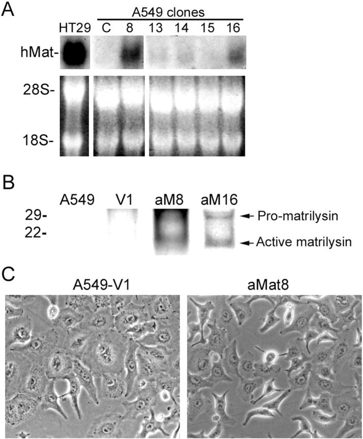
Matrilysin (matrix metalloproteinase-7) is highly expressed in lungs of patients with pulmonary fibrosis and other conditions associated with airway and alveolar injury. Although matrilysin is required for closure of epithelial wounds ex vivo, the mechanism of its action in repair is unknown. We demonstrate that matrilysin mediates shedding of E-cadherin ectodomain from injured lung epithelium both in vitro and in vivo. In alveolar-like epithelial cells, transfection of activated matrilysin resulted in shedding of E-cadherin and accelerated cell migration. In vivo, matrilysin co-localized with E-cadherin at the basolateral surfaces of migrating tracheal epithelium, and the reorganization of cell-cell junctions seen in wild-type injured tissue was absent in matrilysin-null samples. E-cadherin ectodomain was shed into the bronchoalveolar lavage fluid of bleomycin-injured wild-type mice, but was not shed in matrilysin-null mice. These findings identify E-cadherin as a novel substrate for matrilysin and indicate that shedding of E-cadherin ectodomain is required for epithelial repair.


Figures

Figure 1.
Figure 1.
Matrilysin expression in injured human alveolar epithelium. Sections of human lung specimens were stained for matrilysin protein (peroxidase). A: Matrilysin protein was not detected in alveolar epithelium in normal lung. B: Specimen from emphysema with interstitial fibrosis shows intense matrilysin staining in alveolar type II epithelial cells (arrows), particularly bordering sites of epithelial desquamation (arrowheads). Specimens of desquamative interstitial pneumonitis (C) and ARDS (E) show matrilysin staining localized to alveolar type II cells (arrows). Interstitial cells and inflammatory cells were negative for matrilysin staining. D: Serial section of lung from ARDS specimen incubated with preimmune serum. Scale bars, 50 μm.
Figure 2.
Figure 2.
Activated-matrilysin expression in A549 lung epithelial cells. Autoactivating human matrilysin cDNA was stably expressed in A549 cells. A: Northern analysis showed that control cells (C) did not express matrilysin mRNA. Clones 8 and 16 expressed high levels of matrilysin mRNA, whereas clones 13, 14, and 15 expressed low levels. HT29 colon carcinoma cells, which constitutively express matrilysin mRNA, were used as a positive control. The ethidium bromide-stained membrane shows equivalent loading and transfer. B: Matrilysin mRNA-positive clones 8 and 16 were labeled with [35S]-l-methionine-l-cysteine, and secreted matrilysin was immunoprecipitated from conditioned medium. Clones aM8 and aM16 secreted matrilysin and both 29-kd promatrilysin and 19-kd active matrilysin forms were detected. C: Compared to vector-transfected cells (A549-V1), activated-matrilysin-expressing cells (aMat8) were more spindle-shaped and detached from each other.
Figure 3.
Figure 3.
Matrilysin-mediated E-cadherin ectodomain shedding in vitro. A: Immunofluorescence with a monoclonal antibody specific for the human E-cadherin ectodomain showed that E-cadherin localized to cell-cell junctions in vector-transfected cells (A549-V1) but was redistributed to the cytoplasm in aMat8 cells, correlating with decreased cell-cell attachment. B: Western blot for E-cadherin ectodomain (E-cad ecto) showed a reduction in intensity of band for 120-kd full-length E-cadherin in the lysate of aMat expressing cells. C: Increased shedding of an ∼80-kd E-cad ecto-reactive band into the medium (arrowhead). Actin is shown as protein loading control for cell lysates. Calu3 lysate is shown as a control for full-length E-cadherin (arrow). D: Treatment of cells with the hydroxamate MMP inhibitors, SC68180A and RS101625 (MMP-7-specific), inhibited the shedding of the 80-kd fragment into the medium. E: Western blotting of cell lysates with antibody specific for the cytoplasmic domain of human E-cadherin identified an ∼38-kd fragment (arrowhead) in the cell lysate of aMat-expressing cells that was reduced by treatment with SC68180A and was not seen in vector-transfected cells (V1). Full-length E-cadherin is indicated by arrow. Original magnification, ×400 (A).
Figure 4.
Figure 4.
Effect of activated-matrilysin expression on migration. A: Uniform 1-mm wounds were made in confluent aMat-transfected (aMat8) and vector-transfected cells (A549-V1), and wounds were allowed to heal by migration. aMat8 cells closed wounds faster than vector-transfected cells. B: Wound area was calculated at 24 hours and 48 hours after wounding in the presence or absence of 250 μmol/L of hydroxyurea (H). Data represent the average of four experiments ± SD. C: Cells at the wound edge of aMat8 cells were detached (small arrowheads) from the migrating front, whereas control cells (V1) migrated as an intact sheet.
Figure 5.
Figure 5.
Matrilysin co-localization with E-cadherin in migrating tracheal epithelium. Tracheal explants from wild-type C57BL6 mice were wounded, and confocal laser-scanning immunofluorescence microscopy superimposed with differential interference contrast images localized matrilysin and E-cadherin at 0 hours and 24 hours after wounding. A: Unwounded tracheal explant stained with anti-mouse matrilysin antibody and Alexa 568-conjugated secondary antibody. Cut edge of trachea is marked by arrow. B and G: Tracheal explant at 24 hours after wounding showed matrilysin expression (red) at basolateral surfaces of cells just behind leading edge of migrating epithelial front (small arrows). Large arrowhead indicates cilia. C and H: Unwounded trachea stained with anti-mouse E-cadherin ectodomain antibody and Alexa 488-conjugated secondary antibody. E-cadherin expression (green) was present at basolateral surfaces of tracheal epithelium with intense staining at apicolateral adherens junctions (small arrows). D: At 24 hours after wounding, E-cadherin expression remained apicolateral at cells back from wound edge (small arrow), was decreased in cells just behind wound edge (asterisk), and was redistributed evenly around surfaces in cells at leading edge of migrating epithelium (large arrowhead). E: Light microscopic image showed flattened migrating epithelial cell front with loss of cilia. Cells back from cut edge of trachea (arrow) retained cilia (large arrowhead). F: In merged images, matrilysin and E-cadherin were co-localized at basolateral surfaces of cells just behind leading edge of migrating cell front and at the apicolateral surfaces of ciliated cells. I: Close-up of inset in F showing co-localization of matrilysin and E-cadherin (arrows). Original magnifications, ×400.
Figure 6.
Figure 6.
Matrilysin-mediated E-cadherin shedding from wounded tracheal epithelium. Tissue lysates from wild-type and matrilysin-null mice and conditioned media (CM) collected at 24 hours from intact and wounded tracheal explant cultures were analyzed by Western blot with antibody targeted to E-cadherin ectodomain. Equivalent expression of full-length 120-kd E-cadherin is seen in intact tracheal tissue from wild-type (WT) and matrilysin-null (Mat−/−) mice. Immunoreactivity for 80-kd E-cadherin ectodomain is prominently increased in conditioned medium from wounded wild-type tracheas but not in medium from wounded matrilysin-null tracheas.
Figure 7.
Figure 7.
Reorganization of cell-cell contacts in migrating tracheal epithelium. Transmission electron microscopic analysis of wild-type and matrilysin-null tracheal explant cultures at times 0 hours and 6 hours after wounding (uranyl acetate and lead citrate). A and B: Unwounded wild-type trachea showed basolateral membrane interdigitations (arrows). C: By 6 hours after wounding, basolateral interdigitations are lost (arrowheads) from cells at epithelial wound edge of wild-type trachea. D: Basolateral interdigitations persisted (arrows) in cells at wound edge of matrilysin-null trachea. E and F: Higher magnification views of areas indicated by rectangles in C and D. Original magnifications: ×3500 (A); ×10,000 (B); ×7100 (C, D); ×12,500 (E, F).
Figure 8.
Figure 8.
Transmission electron microscopic analysis of wild-type and matrilysin-null tracheal explants at 12 hours after wounding. A: Tracheal epithelial front in wild-type trachea was flattened, and migrating cells spread over basement membrane (BM) into wound. B: Tracheal epithelium in matrilysin-null trachea retained thickness of uninjured epithelial and only cells at very front edge of wound began to spread. C and D: Higher magnification views of areas indicated by rectangles in A and B showed reorganization of cell-cell contacts in wild-type trachea (arrowheads), and persistent membrane interdigitations in matrilysin-null trachea (arrows). Original magnifications: ×3500 (A); ×4500 (B); ×8500 (C); ×11,000 (D).
Figure 9.
Figure 9.
Matrilysin mediates E-cadherin ectodomain shedding in vivo. Wild-type and matrilysin-null mice were instilled with 0.08 U of bleomycin, and bronchoalveolar lavage (BAL) fluid and lung tissue were harvested 3 to 15 days later. A: Western blot for E-cadherin in BAL fluid showed no significant difference in E-cadherin ectodomain immunoreactivity in the first 3 days after bleomycin treatment. At 10 and 15 days, E-cadherin immunoreactivity is increased in BAL fluid from wild-type mice but is not seen in BAL fluid from matrilysin-null mice. B: E-cadherin ectodomain is shed into BAL fluid of bleomycin-treated MMP-9 null and MMP-12 null mice. C: E-cadherin ectodomain immunofluorescence was reduced in alveolar epithelium in lesional areas of bleomycin-injured wild-type mice compared to that in matrilysin-null mice. Signal for E-cadherin is prominent in alveolar epithelium in nonlesional areas of both wild-type and matrilysin-null mice. D: At 10 days after bleomycin, immunofluorescence for E-cadherin and matrilysin were co-localized in damaged alveolar epithelium of wild-type mice (arrows). Original magnifications: ×200 (B); 400 (D).

Similar articles

Cited by

References

    1. Dunnill MS, Massarella GA, Anderson JA: A comparison of the quantitative anatomy of the bronchi in normal subjects, in status asthmaticus, in chronic bronchitis, and in emphysema. Thorax 1969, 24:176-179 - PMC - PubMed
    1. Erjefalt JS, Persson CGA: Airway epithelial repair: breathtakingly quick and multipotentially pathogenic. Thorax 1997, 52:1010-1012 - PMC - PubMed
    1. Laitinen LA, Heino M, Laitinin A, Kava T, Haahtela T: Damage of the airway epithelium and bronchial reactivity in patients with asthma. Am Rev Respir Dis 1985, 131:599-606 - PubMed
    1. Jeffery PK, Wardlaw AJ, Nelson FC, Collins JV, Kay AB: Bronchial biopsies in asthma. An ultrastructural, quantitative study and correlation with hyperreactivity. Am Rev Respir Dis 1989, 140:1745-1753 - PubMed
    1. Myers JL, Katzenstein AL: Ultrastructural evidence of alveolar epithelial injury in idiopathic bronchiolitis obliterans-organizing pneumonia. Am J Pathol 1988, 132:102-109 - PMC - PubMed

Publication types

MeSH terms

LinkOut - more resources