Comparative Study
. 2003 Jun;162(6):1881-93.
doi: 10.1016/S0002-9440(10)64322-2.

Differential expression of the angiogenic factor genes vascular endothelial growth factor (VEGF) and endocrine gland-derived VEGF in normal and polycystic human ovaries

Affiliations
Comparative Study

Differential expression of the angiogenic factor genes vascular endothelial growth factor (VEGF) and endocrine gland-derived VEGF in normal and polycystic human ovaries

Napoleone Ferrara et al. Am J Pathol. 2003 Jun.

Abstract

Angiogenesis is a key aspect of the dynamic changes occurring during the normal ovarian cycle. Hyperplasia and hypervascularity of the ovarian theca interna and stroma are also prominent features of the polycystic ovary syndrome (PCOS), a leading cause of infertility. Compelling evidence indicated that vascular endothelial growth factor (VEGF) is a key mediator of the cyclical corpus luteum angiogenesis. However, the nature of the factor(s) that mediate angiogenesis in PCOS is less clearly understood. Endocrine gland-derived (EG)-VEGF has been recently identified as an endothelial cell mitogen with selectivity for the endothelium of steroidogenic glands and is expressed in normal human ovaries. In the present study, we compared the expression of EG-VEGF and VEGF mRNA in a series of 13 human PCOS and 13 normal ovary specimens by in situ hybridization. EG-VEGF expression in normal ovaries is dynamic and generally complementary to VEGF expression in both follicles and corpora lutea. A particularly high expression of EG-VEGF was detected in the Leydig-like hilus cells found in the highly vascularized ovarian hilus. In PCOS ovaries, we found strong expression of EG-VEGF mRNA in theca interna and stroma in most of the specimens examined, thus spatially related to the new blood vessels. In contrast, VEGF mRNA expression was most consistently associated with the granulosa cell layer and sometimes the theca, but rarely with the stroma. These findings indicate that both EG-VEGF and VEGF are expressed in PCOS ovaries, but in different cell types at different stages of differentiation, thus suggesting complementary functions for the two factors in angiogenesis and possibly cyst formation.


Figures

Figure 1.
Figure 1.
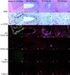
VEGF and EG-VEGF expression in maturing follicles in normal ovaries. A–C: Primary and primordial follicles show strong expression of EG-VEGF (B) but little or no expression of VEGF (C). D–F: Maturing secondary follicles with multiple layers of granulosa cells maintain strong EG-VEGF expression, but show weak to moderate VEGF expression. G–I: Antral follicle (see arrowhead in Figure 5B ▶ ), with abundant mitotic figures (not shown) in both the granulosa and thecal layers, has minimum EG-VEGF expression surrounding the theca, but very intense VEGF expression in the granulosa cell layer and moderate VEGF expression (I) in the thecal cells. J–L: Antral follicle (see filled arrowhead in Figure 4B ▶ ) with heterogeneous EG-VEGF (K) and VEGF (L) expression; the right end of this follicle has a narrow rim of granulosa cells, some of which are degenerating and detached from the theca; these granulosa cells and the surrounding theca externa, lack the significant VEGF expression (L) seen elsewhere in the follicle; adjacent to the area of weak VEGF expression, EG-VEGF thecal expression is focally strong (K). M–O: Mature atretic follicle (see arrow in Figure 4B ▶ ) shows strong expression of EG-VEGF (N) in residual theca interna cells surrounding the glassy membrane (arrows) remnant of the follicular basal lamina. There is weak VEGF expression (O) in a subset of these cells. Scale bars: 100 μm (A–C); 50 μm (D–F); 200 μm (G–O).
Figure 2.
Figure 2.
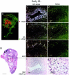
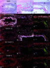
EG-VEGF and VEGF expression in normal ovary early-stage CL. An early-stage (approximately day 2 to 3 after ovulation) CL, characterized by incompletely developed vascularity in the granulosa lutein layer and by inapparent theca lutein cell differentiation (I, J), shows strong VEGF expression in the granulosa lutein cells. A: False-colored autoradiographic film results show intense VEGF expression (red) in the wall of the large cystic CL (B, arrow). Microscopic results show granulosa lutein cells are intensely VEGF-positive (C, dark field; J, bright field), but only weakly positive for EG-VEGF (E); the surrounding theca is only weakly positive for both VEGF and EG-VEGF. VEGFR-2 (KDR) expression (G) is present in small vessels at the boundary between the theca interna and granulosa cell layer, and in vessels invading the outermost granulosa cell layers (I, arrows). Other atretic follicles (A, B) with (closed arrowheads) and without (open arrowhead) intact granulosa cell linings (detail not shown) show prominent EG-VEGF expression in the theca interna. Scale bars: 5 mm (B); 100 μm (C–I); 50 μm (J).
Figure 3.
Figure 3.
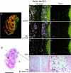
EG-VEGF and VEGF expression in normal ovary early- to mid-stage CL. A CL at approximately day 5 after ovulation, characterized microscopically by well-developed vascularity and early theca lutein cell differentiation (I), shows strong VEGF (C) and EG-VEGF (E) expression in spatially overlapping CL cell populations. A: False-colored autoradiographic film results show yellow-orange overlapping of VEGF (red) and EG-VEGF (green) expression in a large cystic CL. It was not possible to determine from this result whether VEGF and EG-VEGF were co-expressed in the same cells, or in separate cells in the CL. The distribution of EG-VEGF and VEGF signal across the entire CL is consistent with expression by theca granulosa cells, but theca lutein cells may also express EG-VEGF at this stage. Vascular VEGFR-2 (KDR) expression is intense in the CL (G). Scale bars: 5 mm (B); 100 μm (C–I); 50 μm (J).
Figure 4.
Figure 4.
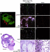
EG-VEGF and VEGF expression in normal ovary mid-stage CL. A: False-colored autoradiographic film results show intense EG-VEGF (green) expression in a narrow convoluted border surrounding a large CL. This mid-stage CL (approximately day 8 after ovulation), characterized by well-developed granulosa lutein vascularity (G) and distinct theca lutein cell differentiation (I), shows intense EG-VEGF expression in the theca lutein cell population at the CL perimeter, surrounding the vessels supplying the CL (E). EG-VEGF and VEGF expression (C) are weak or absent in the granulosa lutein cell layer. Note that VEGF is clearly expressed in this sample in the vascular smooth muscle of some small arterioles supplying the CL (arrowheads, C). VEGFR-2 (KDR) expression is still strong in vessels in all layers of the CL (G). Scale bars: 5 mm (B); 100 μm (C–I); 50 μm (J). GL, granulosa lutein; TL, theca lutein.
Figure 5.
Figure 5.
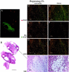
EG-VEGF and VEGF expression in normal ovary late-regressing CL. A regressing CL (approximately day 14 after ovulation), characterized by large, pale, vacuolated theca granulosa and theca lutein cells (I, J), shows absence of both VEGF (C) and EG-VEGF (E) expression. A: False-colored autoradiographic film results show absence of VEGF (red) and EG-VEGF (green) signal in an area that microscopically corresponds to the regressing CL. Only weak VEGFR-2 (KDR) expression (G) is noted in scattered vessels in the granulosa cell layer. A developing tertiary (antral) follicle (A and B, arrowhead) shows strong VEGF expression (see Figure 1 ▶ for details). Scale bars: 5 mm (B); 100 μm (C–I); 50 μm (J).
Figure 6.
Figure 6.
EG-VEGF expression in ovarian hilus cells. EG-VEGF expression (D–F) is very strong in cells, morphologically and biochemically similar to testicular Leydig cells, found in the ovarian hilus in association with blood vessels and unmyelinated nerves. A, D, G: H&E-stained section (A) shows a nest of hilus cells (small arrow) adjacent to a large vein (open arrowheads). In this example, an adjacent unmyelinated nerve fiber (filled arrowhead) lacks associated hilus cells. B, C, E, F: Hilus cells are intimately associated with a large unmyelinated nerve fiber in this section. Epithelium of the rete ovarii (glandular structures in A, top right) expresses neither VEGF nor EG-VEGF. Scale bars: 100 μm (A, B, D, E, G, H); 25 μm (C, F, I).
Figure 7.
Figure 7.
Differential expression of EG-VEGF (green) and VEGF (red) in four individual PCOS specimens. A–D: Parallel tissue sections were hybridized with [33P]-labeled probes, autoradiographic films were digitized, overlaid, and false-colored as described in Materials and Methods. H&E-stained sections (E–H) are also shown. Note the generally nonoverlapping expression pattern of each gene, with VEGF having a more restricted expression, most consistently associated with the lining of several individual cysts. EG-VEGF is expressed around a minority of VEGF-positive (A, B) and around all VEGF-negative cysts, as well as extensively in the stroma. Weak VEGF expression is noted in the fallopian tube mucosa (C, G), where EG-VEGF expression is absent. Scale bar, 5 mm.
Figure 8.
Figure 8.
Distribution of VEGF and EG-VEGF mRNA in parallel sections of cysts in individual PCOS ovaries. Parallel section were hybridized with EG-VEGF anti-sense (D–F), VEGF anti-sense (G–I), EG-VEGF sense (J–L), and VEGF sense (M–O) riboprobes. H&E images (A–C) are shown for reference. A, D, G, J, M: Detail of late-stage atretic follicle from Figure 7A ▶ , boxed area 1; EG-VEGF (D) is strongly expressed in theca cells surrounding follicle lumen in which the granulosa cell layer has degenerated. B, E, H, K, N: Detail of early-stage atretic follicle from Figure 7A ▶ , boxed area 2; VEGF (H) is strongly expressed in granulosa cells surrounding the follicle lumen; some surrounding thecal cells are weakly VEGF-positive; EG-VEGF (C) is expressed in clusters of surrounding thecal cells. C, F, I, L, O: detail from Figure 7D ▶ , boxed area, of two atretic follicles at different stages of degeneration; VEGF (I) is strongly expressed in granulosa cells surrounding lower follicle lumen; the surrounding thin layer of thecal cells are weakly VEGF-positive, and EG-VEGF-negative; EG-VEGF (F) is expressed in the thecal cells of the upper follicle in which the granulosa cell layer has degenerated. Granulosa cells (GC), theca (Th), stroma (St), lumen (L). Scale bars: 200 μm (A, D, G, J, M); 100 μm (B, C, E, F, H, I, K, L, N, O).
Figure 9.
Figure 9.
Correlation between expression of VEGF or EG-VEGF and vascularity, as assessed by expression of CD34, in representative PCOS specimens. Parallel sections were immunostained with anti-CD34 (QBEnd/10, E–H) or hybridized with EG-VEGF anti-sense (I–L), VEGF anti-sense (M–P), EG-VEGF sense (Q–T), and VEGF sense (U–X) riboprobes. H&E images (A–D) are shown for reference. In PCOS ovaries, EG-VEGF expression is high in the theca surrounding atretic follicle lumens (A, B, I, J) and diffusely in ovarian stroma (C, D, K, L), whereas VEGF expression in these areas (Q–T) is weak or absent. Vascularity in corresponding areas is illustrated by CD34 immunostaining (E–H). Similar, although weaker immunostaining was observed with anti-CD31 monoclonal antibody JC/70A (not shown). Scale bars, 100 μm.

Similar articles

Cited by

References

    1. Bassett DL: The changes in the vascular pattern of the ovary of the albino rat during the estrous cycle. Am J Anat 1943, 73:251-278
    1. Zeleznik AJ, Schuler HM, Reichert LE, Jr: Gonadotropin-binding sites in the rhesus monkey ovary: role of the vasculature in the selective distribution of human chorionic gonadotropin to the preovulatory follicle. Endocrinology 1981, 109:356-362 - PubMed
    1. Carr BR, MacDonald PC, Simpson ER: The role of lipoproteins in the regulation of progesterone secretion by the corpus luteum. Fertil Steril 1981, 38:303-311 - PubMed
    1. Goede V, Schmidt T, Kimmina S, Kozian D, Augustin HG: Analysis of blood vessel maturation processes during cyclic ovarian angiogenesis. Lab Invest 1998, 78:1385-1394 - PubMed
    1. Maisonpierre PC, Suri C, Jones PF, Bartunkova S, Wiegend SJ, Radziejewski C, Compton D, McClain J, Aldrich TH, Papadopulos N, Daly TJ, Davis S, Sato TN, Yancopoulos GD: Angiopoietin-2, a natural antagonist for Tie-2 that disrupts in vivo angiogenesis. Science 1997, 277:55-60 - PubMed

Publication types

MeSH terms

Substances