. 2003 Jun;162(6):1913-26.
doi: 10.1016/S0002-9440(10)64325-8.

Distinct role of fibroblast growth factor-2 and vascular endothelial growth factor on tumor growth and angiogenesis

Affiliations

Distinct role of fibroblast growth factor-2 and vascular endothelial growth factor on tumor growth and angiogenesis

Raffaella Giavazzi et al. Am J Pathol. 2003 Jun.

Abstract

Tumors express more than a single angiogenic growth factor. To investigate the relative impact of fibroblast growth factor-2 (FGF-2) and vascular endothelial growth factor (VEGF) on tumor growth and neovascularization, we generated tumor cell transfectants differing for VEGF and/or FGF-2 expression. Human endometrial adenocarcinoma HEC-1-B-derived Tet-FGF-2 cells that express FGF-2 under the control of the tetracycline-responsive promoter (Tet-off system) were further transfected with a VEGF(121) anti-sense (AS-VEGF) cDNA. Next, Tet-FGF-2 and AS-VEGF/Tet-FGF-2 cells were transplanted subcutaneously in nude mice that received tetracycline or not in the drinking water. Simultaneous expression of FGF-2 and VEGF in Tet-FGF-2 cells resulted in fast-growing lesions characterized by high blood vessel density, patency and permeability, and limited necrosis. Blood vessels were highly heterogeneous in size and frequently associated with pericytes. Inhibition of FGF-2 production by tetracycline caused a significant decrease in tumor burden paralleled by a decrease in blood vessel density and size. AS-VEGF expression resulted in a similar reduction in blood vessel density associated with a significant decrease in pericyte organization, vascular patency, and permeability. The consequent decrease in tumor burden was paralleled by increased tumor hypoxia and necrosis. A limited additional inhibitory effect was exerted by simultaneous down-regulation of FGF-2 and VEGF expression. These findings demonstrate that FGF-2 and VEGF stimulate vascularization synergistically but with distinctive effects on vessel functionality and tumor survival. Blockade of either one of the two growth factors results in a decrease in blood vessel density and, consequently, in tumor burden. However, inhibition of the expression of VEGF, but not of FGF-2, affects also vessel maturation and functionality, leading to tumor hypoxia and necrosis. Our experimental model represents an unique tool to investigate anti-neoplastic therapies in different angiogenic environments.


Figures

Figure 1.
Figure 1.
VEGF and FGF-2 expression by Tet-FGF-2 and AS-VEGF/Tet-FGF-2 transfectants. Tet-FGF-2 15H cells were transfected with the pcDNA-3 expression vector harboring the VEGF121 anti-sense cDNA. A: Stable transfectants (AS-VEGF/Tet-FGF-2 A5 and C1 clones) and mock-transfected cells (Tet-FGF-2 A4 and C7 clones) were analyzed for AS-VEGF mRNA steady-state levels by Northern blotting of total RNA (20 μg/lane) using a digoxigenin-UTP-labeled AS-VEGF riboprobe. B: Uniform loading of the gel was assessed by methylene blue staining of the filter. C: Serum-free conditioned media from the different clones (60 μg of protein) were probed with anti-VEGF antibodies by Western blotting showing decreased levels of the secreted forms of VEGF in AS-VEGF/Tet-FGF-2 cells. D: Lysates (500 μg) from cells grown in the absence (−) or in the presence (+) of tetracycline were loaded onto 0.1-ml heparin-Sepharose columns and bound material was probed with anti-FGF-2 antibodies in a Western blot. All of the clones express the 24-, 22-, and 18-kd FGF-2 isoforms whose expression is hampered by tetracycline treatment. E: Endothelial GM 7373 cells were incubated for 24 hours with increasing concentrations of the conditioned media from untreated and tetracycline-treated (+tet) Tet-FGF-2 A4 and AS-VEGF/Tet-FGF-2 C1 clones. At the end of incubation cells were trypsinized and counted. Data are the mean ± SE of four determinations.
Figure 2.
Figure 2.
In vitro and in vivo growth properties of Tet-FGF-2 and AS-VEGF/Tet-FGF-2 transfectants. A: AS-VEGF/Tet-FGF-2 A5 and C1 clones (▵, ▴) and mock Tet-FGF-2 C7 and A4 clones (○, •) were seeded at 25,000 cells/well in 24-well plates in complete cell culture medium. At the indicated time points, cells were trypsinized and counted. B: Cells were transplanted subcutaneously in nude mice (1 × 10⁶ cells/animal) and tumor growth was reported. Each point is the mean ± SE of five tumors. Statistical analysis of tumor volume by orthogonal comparison analysis of variance at day 45 showed a significant difference (P < 0.001) between Tet-FGF-2 and AS-VEGF/Tet-FGF-2 clones (**). At sacrifice, three Tet-FGF-2 A4 tumors and three AS-VEGF/Tet-FGF-2 C1 tumors were analyzed for AS-VEGF mRNA expression (arrow) by Northern blotting (inset). Uniform loading of the gel was assessed by methylene blue staining of the filter.
Figure 3.
Figure 3.
Effect of tetracycline on Tet-FGF-2 and AS-VEGF/Tet-FGF-2 tumor growth. Nude mice were transplanted subcutaneously with 1 × 10⁶ Tet-FGF-2 A4 cells (○, •) or AS-VEGF/Tet-FGF-2 C1 cells (▵, ▴). Animals were left untreated (•, ▴) or received 2 mg/ml of tetracycline in the drinking water throughout the whole experimental period starting 4 days before cell transplantation (○, ▵). Each point is the mean ± SE of five tumors. The results are representative of three independent experiments. Statistical analysis of tumor volumes by analysis of variance followed by Tukey-Kramer posthoc test showed a significant difference (P < 0.01) between the untreated Tet-FGF-2 group and all of the other groups (day 43, **) and between tetracycline-treated Tet-FGF-2 group and the two AS-VEGF/Tet-FGF-2 groups (day 43 and day 64, *). No significant difference was observed between untreated and tetracycline-treated AS-VEGF/Tet-FGF-2 groups at any time point.
Figure 4.
Figure 4.
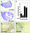
Tumor necrosis and mononuclear cell infiltrate in Tet-FGF-2 and AS-VEGF/Tet-FGF-2 xenografts. At the end of the experimentation (shown in Figure 3 ▶ ), animals were sacrificed and tumor sections were stained with H&E (A, B). Note the large necrotic area at the center of AS-VEGF/Tet-FGF-2 lesions (arrows in B) compared to the limited necrosis observed in Tet-FGF-2 tumors (A). In C, the percentage of necrotic tumor parenchyma was quantified by computerized image analysis of tissue sections (two slides per tumor, five animals per group) from Tet-FGF-2 and AS-VEGF/Tet-FGF-2 xenografts treated (+) or not (−) with tetracycline. **, Statistically different from AS-VEGF (P < 0.05 or better). D and E: CD11b immunostaining of Tet-FGF-2 and AS-VEGF/Tet-FGF-2 tumors. Large areas of CD11b+ monocyte/macrophage infiltrates are observed in Tet-FGF-2 xenografts (D) but not in AS-VEGF/Tet-FGF-2 lesions (E). A similar immunostaining was observed in all tumors examined (five tumors per group). Original magnifications: ×2.5 (A, B); ×20 (D, E); ×63 (inset in D).
Figure 5.
Figure 5.
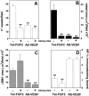
Vascularization of Tet-FGF-2 and AS-VEGF/Tet-FGF-2 tumors. Tet-FGF-2 and AS-VEGF/Tet-FGF-2 tumor sections from control (−) and tetracycline-treated (+) nude mice (see Figure 3 ▶ ) were processed for CD31 immunostaining. For each section, the most vascularized area within the tumor parenchyma at the periphery of the lesion was selected and microvessels in a ×400 field were counted (A). Total CD31+ blood vessel area (B) and total pericyte/myofibroblast α-SMA+ area (C) in 0.4-mm2 fields of viable tumor parenchyma (three fields per tumor) were quantified by computerized image analysis. D: Semiquantitative scoring of HIF-1α immunostaining was performed on the whole tumor sections using an arbitrary scale from 0 (negative) to 4+ (strongly positive). n = 5 mice per group. **, P < 0.05 or better.
Figure 6.
Figure 6.
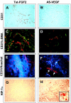
Immunohistochemical characterization of Tet-FGF-2 and AS-VEGF/Tet-FGF-2 tumors from tetracycline-untreated mice. Representative images from Tet-FGF-2 (A, C, E, G) and AS-VEGF/Tet-FGF-2 (B, D, F, H) lesions are shown. See Figure 5 ▶ for quantitative analysis performed on both tumor types in the absence and in the presence of tetracycline treatment. A and B: Tumor sections were processed for CD31 immunostaining. Note the large-sized vessels in Tet-FGF-2 tumors (A) when compared to small-caliber microvessels in AS-VEGF/Tet-FGF-2 lesions (B). Small-caliber microvessels were observed also in all of the Tet-FGF-2 and AS-VEGF/Tet-FGF-2 tumors from tetracycline-treated animals (not shown). C and D: Tumor sections were double-labeling immunostained to visualize CD31+ endothelial cells (in red) and α-SMA+ cells (in green) by confocal microscopy. Note the numerous α-SMA+ cells frequently associated with CD31+ blood vessels in Tet-FGF-2 lesions (C) when compared to AS-VEGF/Tet-FGF-2 tumors (D). E and F: Animals were injected intravenously 2 minutes before sacrifice with fluorescent Hoechst 33342. A remarkable extravasation of the dye (in blue) from patent CD31+ vessels (in red) was observed in Tet-FGF-2 lesions (E) when compared to AS-VEGF/Tet-FGF-2 tumors (F). Note the nonpatent Hoechst 33342-negative/CD31+ vessels frequently detected in AS-VEGF/Tet-FGF-2 lesions (arrows in F). G and H: HIF-1α immunostaining of Tet-FGF-2 (G) and AS-VEGF/Tet-FGF-2 (H) tumors showing a strong, mainly nuclear (inset), HIF-1α expression in AS-VEGF-transfected lesions. Tetracycline administration had no significant effect on α-SMA and HIF-1α immunostaining and on Hoechst 33342 extravasation in both Tet-FGF-2 and AS-VEGF/Tet-FGF-2 tumors when compared to untreated lesions (data not shown). Original magnifications: ×20 (A–H); ×63 (inset in H).

Similar articles

Cited by

References

    1. Folkman J: Angiogenesis in cancer, vascular, rheumatoid and other disease. Nat Med 1995, 1:27-31 - PubMed
    1. Carmeliet P, Jain RK: Angiogenesis in cancer and other diseases. Nature 2000, 407:249-257 - PubMed
    1. Relf M, LeJeune S, Scott PA, Fox S, Smith K, Leek R, Moghaddam A, Whitehouse R, Bicknell R, Harris AL: Expression of the angiogenic factors vascular endothelial cell growth factor, acidic and basic fibroblast growth factor, tumor growth factor beta-1, platelet-derived endothelial cell growth factor, placenta growth factor, and pleiotrophin in human primary breast cancer and its relation to angiogenesis. Cancer Res 1997, 57:963-969 - PubMed
    1. Moscatelli D, Presta M, Rifkin DB: Purification of a factor from human placenta that stimulates capillary endothelial cell protease production, DNA synthesis, and migration. Proc Natl Acad Sci USA 1986, 83:2091-2095 - PMC - PubMed
    1. Shing Y, Folkman J, Sullivan R, Butterfield C, Murray J, Klagsbrun M: Heparin affinity: purification of a tumor-derived capillary endothelial cell growth factor. Science 1984, 223:1296-1299 - PubMed

Publication types

MeSH terms