. 2003 Jun;162(6):1927-36.
doi: 10.1016/S0002-9440(10)64326-X.

Angiogenic actions of angiopoietin-1 require endothelium-derived nitric oxide

Affiliations

Angiogenic actions of angiopoietin-1 require endothelium-derived nitric oxide

Saeid Babaei et al. Am J Pathol. 2003 Jun.

Abstract

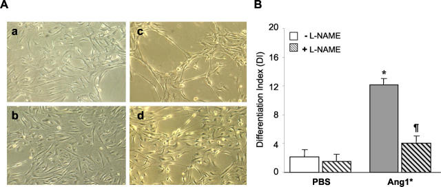
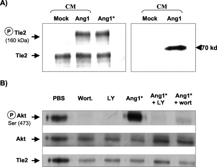
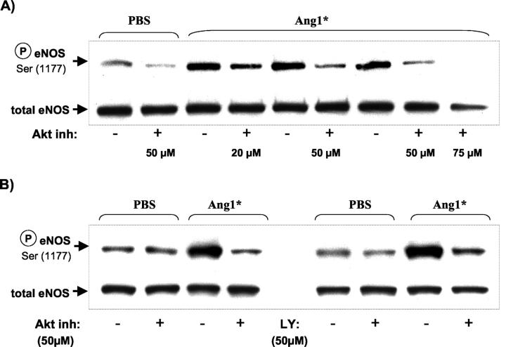
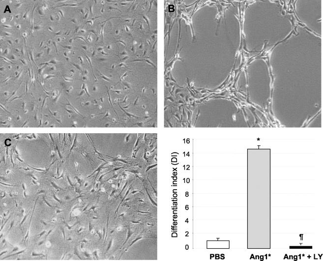
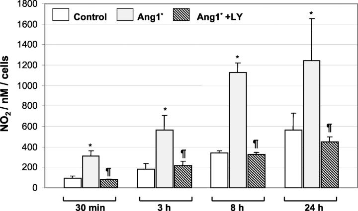
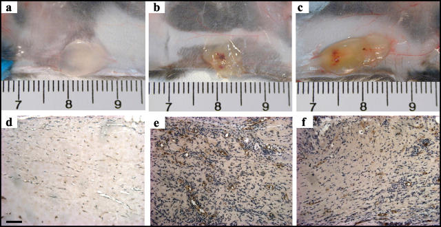
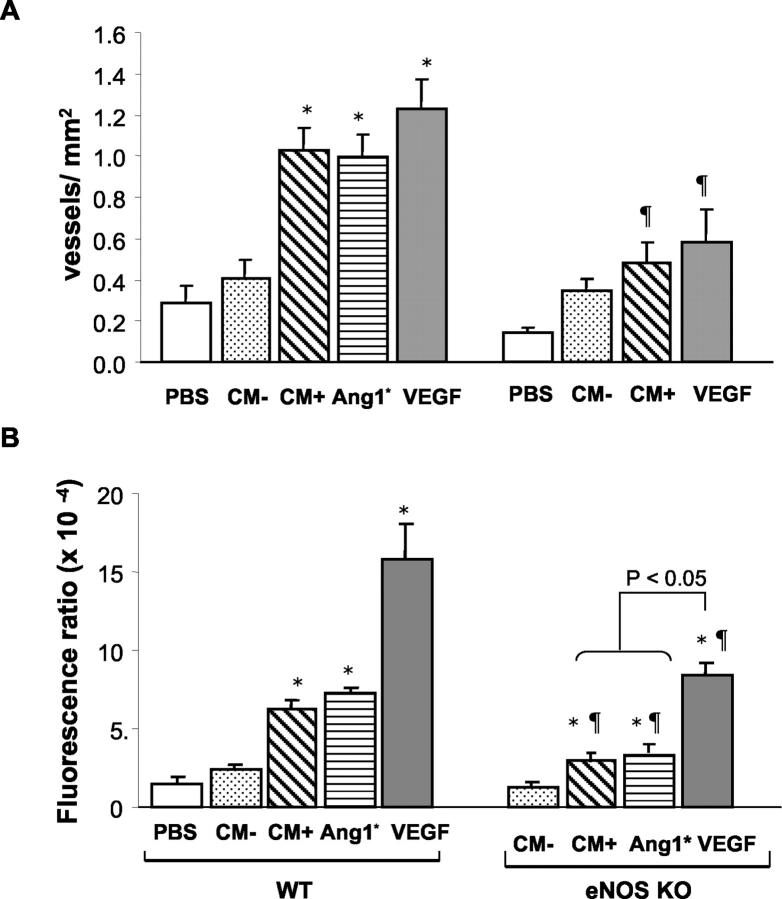
Angiopoietin1 (Ang1) is a novel angiogenic factor with important actions on endothelial cell (EC) differentiation and vascular maturation. Ang1 has been shown to prevent EC apoptosis through activation of PI3-kinase/Akt, a pathway that is also known to activate endothelium nitric oxide synthase (eNOS). Therefore, we hypothesized that the angiogenic effects of Ang1 would also be dependent on the PI3-kinase/Akt pathway, possibly mediated by increased eNOS activity and NO release. Treatment of human umbilical vein endothelial cells with recombinant Ang1* (300 ng/ml) for 15 minutes resulted in PI3-kinase-dependent Akt phosphorylation, comparable to that observed with vascular endothelial growth factor (VEGF) (50 ng/ml), and increased NO production in a PI3-kinase/Akt-dependent manner. Capillary-like tube formation induced by Ang1* in fibrin matrix at 24 hours (differentiation index, DI: 13.74 +/- 0.76 versus control 1.71 +/- 0.31) was abolished in the presence of the selective PI3-kinase inhibitor, LY294002 (50 micro mol/L) (DI: 0.31 +/- 0.31, P < 0.01) or the NOS inhibitor, L-NAME (3 mmol/L) (DI: 4.10 +/- 0.59, P < 0.01). In subcutaneous Matrigel implants in vivo, addition of recombinant Ang1* or wild-type Ang1 from conditioned media of COS-1 cells transfected with a pFLAG Ang1 expression vector, induced significant neovascularization to a degree similar to VEGF. Finally, angiogenesis in vivo in response to both Ang1 and VEGF was significantly reduced in eNOS-deficient compared with wild-type mice. In summary, our results demonstrate for the first time that endothelial-derived NO is required for Ang1-induced angiogenesis, and that the PI3-kinase signaling mediates the activation of eNOS and NO release in response to Ang1.


Figures

Figure 1.
Figure 1.
Nitric oxide-synthase activity required for angiogenic effects of Ang1. A: Ang1*-induced capillary-like tube formation in the three-dimensional fibrin matrices is blocked by inhibition of endogenous eNOS activity. HUVECs were cultured on fibrin matrices in the presence (c and d) or absence (a and b) of recombinant Ang1*, with L-NAME (b and d) or without L-NAME (a and c) for 24 hours. A significant increase in tube length was observed in the presence of Ang1* which was inhibited by L-NAME (original magnification, ×100). B: Quantitative analysis of capillary-like network formation calculated as the ratio of total tube length over cell area for each field (DI, differentiation index). Scale bars represent a mean ± SEM of DI value obtained for each culture well from six independent experiments. The difference among groups were analyzed by analysis of variance followed by Student’s t-test. *P < 0.05 versus control, #P < 0.05 versus Ang1*. Ang1* (300 ng/ml), L-NAME (3 mmol/L).
Figure 2.
Figure 2.
Ang1 stimulates Tie2 and Akt phosphorylation. A: Tie2 receptor tyrosine phosphorylation by Ang1-CM or Ang1* in HUVECs visualized by immunoblot analysis with mouse monoclonal anti-phosphotyrosine antibodies (left panel, top), and anti-Tie2 antibody to normalize equal loading (bottom). Note that the similar level of phosphorylation of Tie2 could be detected following stimulation with Ang1-CM or Ang1* recombinant protein, whereas no phosphorylation was seen following stimulation of cells with mock-CM. Western blot analysis of the concentrated CM obtained from pFLAG (mock)-transfected COS-1 cells or cells expressing pFLAG-Ang1, shows a single 70-kd band, confirming the expression of Ang1 as a c-Myc fused protein (right). Data shown are representative of two independent experiments. B: Stimulation of HUVEC with Ang1* (300 ng/ml), increased Akt phosphorylation, which was blocked by the PI3-kinase inhibitors, LY294002 (50 μmol/L) or wortmannin (100 nmol/L). All figures are representative of three separate experiments. Akt phosphorylation (P-Akt, Ser 473, top); total Akt expression (center) and total Tie2 expression (bottom).
Figure 3.
Figure 3.
Ang1*-dependent phosphorylation of eNOS is blocked by PI3-kinase and Akt inhibitors. A: HUVEC were maintained overnight in F12 medium with 1% FBS, then pretreated for 2 hours in serum-free medium with an Akt inhibitor at various doses (20 to 75 μmol/L) to evaluate optimal conditions as indicated. B: HUVECs were cultured as above and pretreated either with an Akt inhibitor (50 μmol/L) or a PI3-kinase inhibitor LY294002 (50 μmol/L). In both panels the cells were either stimulated with Ang1* (300 ng/ml, 30 minutes) or buffer solution. After the stimulation, Western analysis was performed using antibodies specific to phosphorylated eNOS and total eNOS.
Figure 4.
Figure 4.
Ang1*-induced activation of PI3-kinase/Akt pathway is required for endothelial cell tube formation. A: HUVEC plated on fibrin matrices were pretreated with 50 μmol/L of LY294002 for 2 hours before addition of Ang1* (300 ng/ml) or PBS, and then incubated for 24 hours. HUVEC grown on fibrin gel in the presence of PBS (control) showed no significant angiogenic response (A). In contrast, cells treated with Ang1* showed an extensive network of capillary-like tube formation (B), which was prevented by addition of LY294002 (C). Original magnification, ×100. Bars represent the mean ± SEM of four independent experiments. Statistical comparisons were performed using one-way analysis of variance followed by Student’s t-test. *P < 0.05 versus control, P < 0.05 versus Ang1*.
Figure 5.
Figure 5.
Ang1* activates NO production and release. HUVEC CM was collected after incubation with Ang1* (300 ng/ml) for the periods indicated, and assayed for NO production, as described in the Methods section. Results are expressed as the mean ± SEM of three independent experiments (five experiments for the 24-hour time point). *P < 0.05 versus PBS, P < 0.05 versus Ang1*.
Figure 6.
Figure 6.
Ang1-induced neovascularization of Matrigel plugs in vivo. Gross appearance of Matrigel plugs containing PBS (a), Ang1* (300 ng/ml, b) or VEGF (100 ng/ml, c) implanted for 10 days in WT mice. Histological sections of the Matrigel plugs retrieved from WT (d and e: mock-CM and Ang1-CM, respectively) or eNOS KO (f: Ang1-CM) mice and immunostained with vWF to identify neovessels.
Figure 7.
Figure 7.
Quantative analysis of neovascularization of the Matrigel plugs at day 10. A: Number of vWF positive vessels per mm2 of Matrigel plugs implanted into WT or eNOS KO mice. B: Blood content in the Matrigel explants assessed by FITC-dextran perfusion in both WT and eNOS KO mice. Data are presented as mean ± SEM (6 animals per group). mock-CM (CM−), Ang1-CM (CM+), Ang1* and VEGF (both 300 ng/ml). *P < 0.05 versus PBS or CM−, P < 0.05 versus WT mice.

Similar articles

Cited by

References

    1. Davis S, Aldrich TH, Jones PF, Acheson A, Compton DL, Jain V, Ryan TE, Bruno J, Radziejewski C, Maisonpierre PC, Yancopoulos GD: Isolation of angiopoietin-1, a ligand for the TIE2 receptor, by secretion-trap expression cloning. Cell 1996, 87:1161-1169 - PubMed
    1. Puri MC, Rossant J, Alitalo K, Bernstein A, Partanen J: The receptor tyrosine kinase TIE is required for integrity and survival of vascular endothelial cells. EMBO J 1995, 14:5884-5891 - PMC - PubMed
    1. Dumont DJ, Gradwohl G, Fong GH, Puri MC, Gertsenstein M, Auerbach A, Breitman ML: Dominant-negative and targeted null mutations in the endothelial receptor tyrosine kinase, tek, reveal a critical role in vasculogenesis of the embryo. Genes Dev 1994, 8:1897-1909 - PubMed
    1. Sato TN, Tozawa Y, Deutsch U, Wolburg-Buchholz K, Fujiwara Y, Gendron-Maguire M, Gridley T, Wolburg H, Risau W, Qin Y: Distinct roles of the receptor tyrosine kinases Tie-1 and Tie-2 in blood vessel formation. Nature 1995, 376:70-74 - PubMed
    1. Suri C, Jones PF, Patan S, Bartunkova S, Maisonpierre PC, Davis S, Sato TN, Yancopoulos GD: Requisite role of angiopoietin-1, a ligand for the TIE2 receptor, during embryonic angiogenesis. Cell 1996, 87:1171-1180 - PubMed

Publication types

MeSH terms

Substances

LinkOut - more resources