. 2003 Jun;162(6):2019-28.
doi: 10.1016/S0002-9440(10)64334-9.

A murine model to study leukocyte rolling and intravascular trafficking in lung microvessels

Affiliations

A murine model to study leukocyte rolling and intravascular trafficking in lung microvessels

Lyudmila Sikora et al. Am J Pathol. 2003 Jun.

Abstract

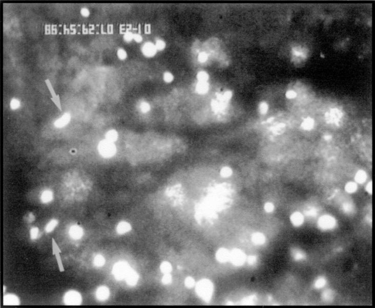
The cascade of leukocyte interactions under conditions of blood flow is well established in the systemic microcirculation, but not in lung microcirculation. We have developed a murine model to study lung microcirculation by transplanting lung tissue into dorsal skin-fold window chambers in nude mice and examining the ability of leukocytes to traffic within revascularized lung microvessels by intravital microscopy. The revascularized lung allograft demonstrated a network of arterioles, capillaries, and postcapillary venules with continuous blood flow. Stimulation of the lung allograft with TNF-alpha induced leukocyte rolling and adhesion in both arterioles and venules. Treatment with function-blocking anti-selectin mAb revealed that P- and L-selectin are the predominant rolling receptors in the lung microvessels, with E-selectin strengthening P-selectin-dependent interactions. Intravital microscopic studies also demonstrated that during their transit in capillaries, some leukocytes undergo shape change and continue to roll as elongated cells in postcapillary venules. Furthermore, the revascularized microvessels demonstrated the ability to undergo vasoconstriction in response to superfusion with endothelin-1. Overall, these studies demonstrate that the revascularized lung allograft is responsive to various external stimuli such as cytokines and vaso-active mediators and serves as a model to evaluate the interaction of leukocytes with the vascular endothelium in the lung microcirculation under acute as well as chronic experimental conditions.


Figures

Figure 1.
Figure 1.
Photomicrograph of lung allograft transplanted into dorsal skin-fold window chamber of a nude mouse. Lung allografts (white arrows) excised from donor neonatal mice are transplanted into the skin-fold window chamber (black arrow) implanted in the dorsal skin of recipient nude mice.
Figure 2.
Figure 2.
Establishment of blood flow in the lung allograft. A: Lung allografts from neonatal mice are labeled with CMTMR (arrows) and implanted into the dorsal skin-fold window chamber of nude mice. Establishment of blood flow on day 0 (B), day 4 (C), and day 9 (D) can be visualized by administration of FITC-dextran 500,000 and stroboscopic epi-illumination. While blood flow is evident in the adjoining host vessels, no flow is apparent within the allograft (arrows) on day 0 (B). The lung allografts revascularize significantly (∼50%) by day 4 (C) and completely by day 9 (D).
Figure 3.
Figure 3.
The lung allograft establishes blood flow by initiating contact with the host vessels. The blood vessels in the periphery of the lung allograft (black arrows), visualized by plasma enhancement with FITC-dextran, appear to make contact by extending into the host tissue. The white background in panels A (lower magnification) and B (higher magnification) represent blood flow in the recipient cutaneous vessels, but not in the donor lung allograft vessels.
Figure 4.
Figure 4.
Photomicrographs of a completely revascularized lung allograft in the skin-fold chamber of nude mice. The implanted lung allograft has established connections with the recipient cutaneous vessels all around the circumference of the allograft (A). The host vessels in the absence of a transplanted allograft are shown in (B).
Figure 5.
Figure 5.
H&E-stained histology of normal mouse skin and the revascularized lung allograft in the dorsal skin-fold chamber of nude mice. A: Section of mouse dorsal skin 14 days after implantation of the titanium chamber showing the characteristic squamous epithelium (black arrow), hair follicles (blue arrow) and sebaceous glands (red arrow); magnification ×400. B: Cross-section of the lung allograft outlined by smooth muscle cells (black arrow) and surrounded by the dorsal skin (blue arrow); magnification ×100. C: Cross-section of the lung allograft showing the bronchus (black arrow) and the pulmonary artery (blue arrow); magnification ×400. D: Cross-section of the lung allograft showing the bronchus lined by bronchial epithelial cells (black arrow) with protruding cilia (blue arrow); magnification ×1000.
Figure 6.
Figure 6.
Effect of ET-1 on lung microvascular vessel diameter. The effect of ET-1 on the microvessel diameter of the revascularized lung allograft was assessed over a period of 45 minutes. Local superfusion of the lung allograft with ET-1 (1.0 μmol/L) but not diluent (PBS) alone (CONTROL) resulted in significant vasoconstriction of the LMV.
Figure 7.
Figure 7.
Effect of TNF-α stimulation on leukocyte rolling (A) and adhesion (B) in arterioles and postcapillary venules in LMV. The lung allograft (day 9) was superfused with TNF-α and the interaction of acridine orange-labeled host leukocytes in arterioles and postcapillary venules was investigated 4 to 6 hours after cytokine stimulation. TNF-α induced significant leukocyte rolling (A) and adhesion (B) in both arterioles and postcapillary venules of the lung allograft. Data represented is mean ± SEM.
Figure 8.
Figure 8.
Leukocytes undergo intravascular shape change in capillaries of the lung allograft. Increased rolling of acridine orange-labeled leukocytes in lung allograft microvessels and retention of trapped leukocytes in the capillary bed for variable periods of time were observed in the presence of TNF-α. A fraction of the leukocytes passing through the capillary bed were observed to stretch and elongate (arrows) before entering the postcapillary venules.
Figure 9.
Figure 9.
Effect of anti-selectin mAbs on TNF-α stimulated leukocyte rolling in LMV. Nude mice were administered with mAbs against leukocyte-expressed L-selectin (L-sel) and endothelial-expressed E-selectin (E-sel) or P-selectin (P-sel) individually (A) as well as a combination of anti-selectin mAbs (P+E-sel, P+L-sel, L+E-sel, P+L+E-sel) (B) and their ability to inhibit leukocyte rolling in TNF-α stimulated LMV was investigated. Data represent normal leukocyte rolling (PBS) as well as after receptor blockade with specific mAb treatment (2 mg/kg body weight). Normal rat IgG was used as a control. Values represent rollers/minute (mean ± SEM) from 286 vessels and 11 animals.

Similar articles

Cited by

References

    1. Kuebler WM, Kuhnle GEH, Groh J, Goetz AE: Leukocyte kinetics in pulmonary microcirculation: intravital fluorescence microscopic study. J Appl Physiol 1994, 76:65-71 - PubMed
    1. Kuhnle GEH, Leipfinger FH, Goetz AE: Measurement of microhemodynamics in the ventilated rabbit lung by intravital fluorescence microscopy. J Appl Physiol 1993, 74:1462-1471 - PubMed
    1. Davis MJ, Gilmore JP, Joyner WL: Responses of pulmonary allograft and cheek pouch arterioles in the hamster to alterations in extravascular pressure in different oxygen environments. Circ Res 1981, 49:133-140 - PubMed
    1. Davis MJ, Joyner WL, Gilmore JP: Microvascular pressure distribution and responses of pulmonary allografts and cheek pouch arterioles in the hamster to oxygen. Circ Res 1981, 49:125-132 - PubMed
    1. Shepard J, Joyner WL, Gilmore JP: Hypoxia does not alter angiotensin converting enzyme activity in hamster pulmonary microvessels. Circ Res 1987, 61:228-235 - PubMed

MeSH terms

LinkOut - more resources