. 2003 Jun;162(6):2029-39.
doi: 10.1016/S0002-9440(10)64335-0.

Absence of caveolin-1 sensitizes mouse skin to carcinogen-induced epidermal hyperplasia and tumor formation

Affiliations

Absence of caveolin-1 sensitizes mouse skin to carcinogen-induced epidermal hyperplasia and tumor formation

Franco Capozza et al. Am J Pathol. 2003 Jun.

Abstract

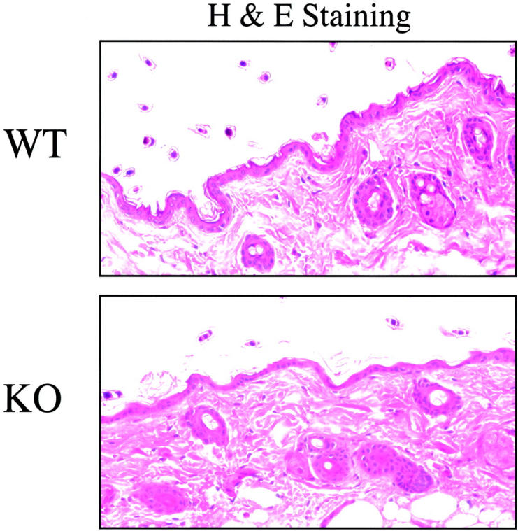
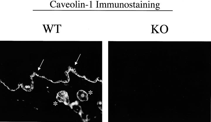
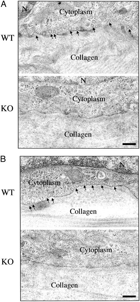
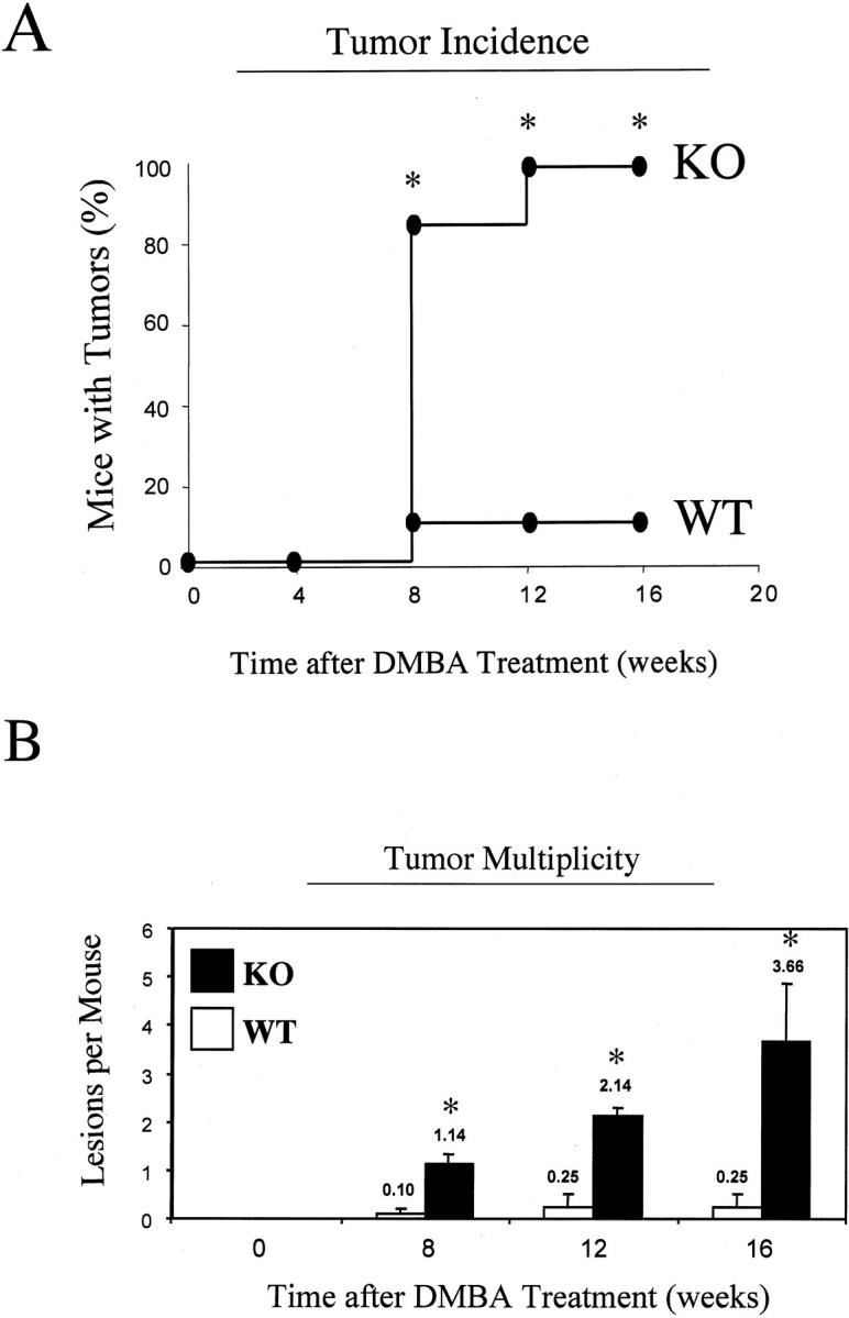
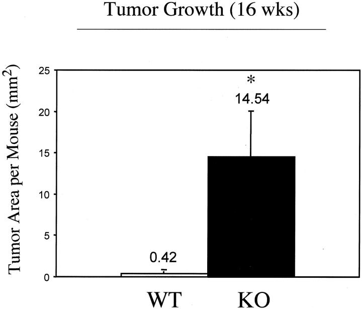
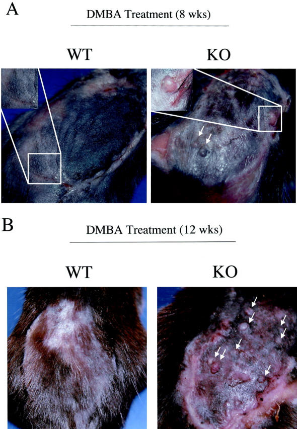
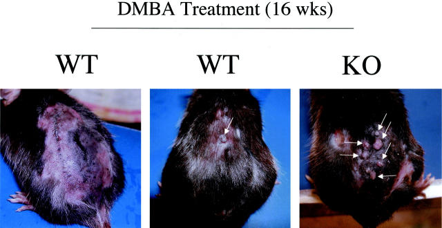
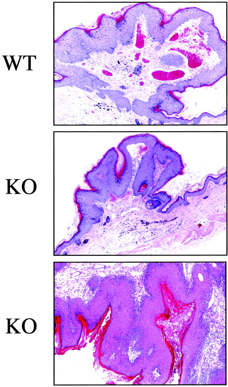
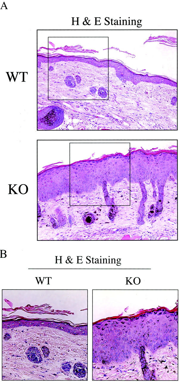
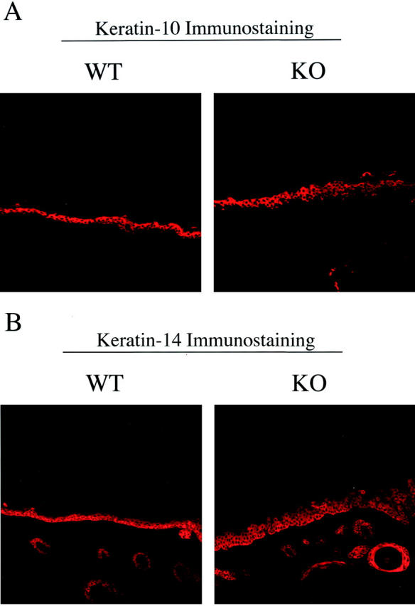
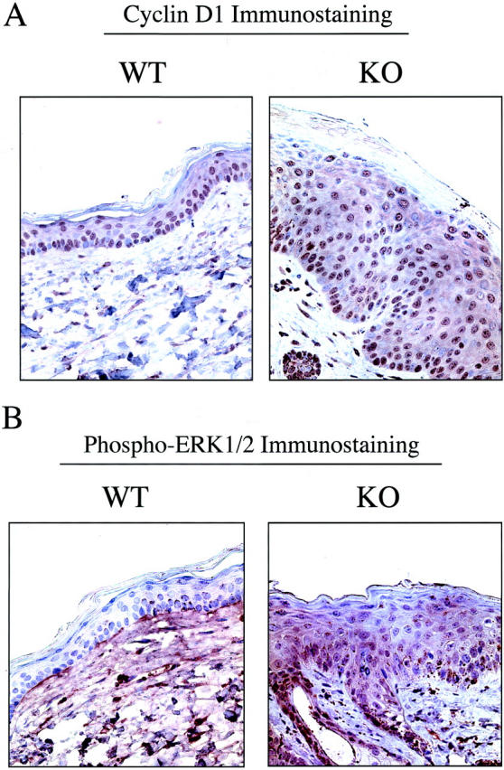
Caveolin-1 is the principal protein component of caveolae membrane domains, which are located at the cell surface in most cell types. Evidence has accumulated suggesting that caveolin-1 may function as a suppressor of cell transformation in cultured cells. The human CAV-1 gene is located at a putative tumor suppressor locus (7q31.1/D7S522) and a known fragile site (FRA7G) that is deleted in a variety of epithelial-derived tumors. Mechanistically, caveolin-1 is known to function as a negative regulator of the Ras-p42/44 MAP kinase cascade and as a transcriptional repressor of cyclin D1, possibly explaining its transformation suppressor activity in cultured cells. However, it remains unknown whether caveolin-1 functions as a tumor suppressor gene in vivo. Here, we examine the tumor suppressor function of caveolin-1 using Cav-1 (-/-) null mice as a model system. Cav-1 null mice and their wild-type counterparts were subjected to carcinogen-induced skin tumorigenesis, using 7,12-dimethylbenzanthracene (DMBA). Mice were monitored weekly for the development of tumors. We demonstrate that Cav-1 null mice are dramatically more susceptible to carcinogen-induced tumorigenesis, as they develop skin tumors at an increased rate. After 16 weeks of DMBA-treatment, Cav-1 null mice showed a 10-fold increase in tumor incidence, a 15-fold increase in tumor number per mouse (multiplicity), and a 35-fold increase in tumor area per mouse, as compared with wild-type littermate mice. Moreover, before the development of tumors, DMBA-treatment induced severe epidermal hyperplasia in Cav-1 null mice. Both the basal cell layer and the suprabasal cell layers were expanded in treated Cav-1 null mice, as evidenced by immunostaining with cell-type specific differentiation markers (keratin-10 and keratin-14). In addition, cyclin D1 and phospho-ERK1/2 levels were up-regulated during epidermal hyperplasia, suggesting a possible mechanism for the increased susceptibility of Cav-1 null mice to tumorigenesis. However, the skin of untreated Cav-1 null mice appeared normal, without any evidence of epidermal hyperplasia, despite the fact that Cav-1 null keratinocytes failed to express caveolin-1 and showed a complete ablation of caveolae formation. Thus, Cav-1 null mice require an appropriate oncogenic stimulus, such as DMBA treatment, to reveal their increased susceptibility toward epidermal hyperplasia and skin tumor formation. Our results provide the first genetic evidence that caveolin-1 indeed functions as a tumor suppressor gene in vivo.


Figures

Figure 1.
Figure 1.
Cav-1 null mice show normal skin morphology. Skin biopsies were taken from the backs of untreated adult wild-type and Cav-1 null mice (4 months old). After excision, samples were formalin-fixed, paraffin-embedded, and stained with H&E. Note that Cav-1 null skin appears normal, as compared with wild-type control mice. Importantly, there is no evidence of epidermal hyperplasia or hyperkeratosis. Images were acquired with a 40× objective. Similar results were obtained with younger mice.
Figure 2.
Figure 2.
Caveolin-1 immunostaining in wild-type and Cav-1 null skin biopsies. Paraffin-embedded sections of skin biopsies derived from the backs of untreated adult wild-type and Cav-1 null mice were immunostained with antibodies directed against the N-terminal domain of caveolin-1 (rabbit pAb, N-20; Santa Cruz Biotech, Inc.). Note that in wild-type mice, caveolin-1 is highly expressed in keratinocyes, especially within the basal cell layer (arrow) and the hair follicles (asterisks). Images shown were acquired with a 20× objective.
Figure 3.
Figure 3.
Cav-1 null keratinocytes lack morphologically identifiable caveolae. Skin biopsies taken from untreated wild-type and Cav-1 null mice were processed for transmission electron microscopy. Note that in wild-type animals, caveolae organelles (50- to 100-nm vesicles and invaginations; arrows) are particularly abundant in keratinocytes within the basal cell layer (A) and the hair follicles (B). Interestingly, the distribution of caveolae in keratinocytes is restricted to the basal membrane, but they are absent from the lateral and apical regions of the plasma membrane. However, in Cav-1 null keratinocytes, note that caveolae formation is completely ablated. The cytoplasm and extracellular collagen fibrils are as indicated. N, nucleus. Scale bar, 250 nm.
Figure 4.
Figure 4.
Cav-1 null mice are dramatically more susceptible to DMBA-induced skin carcinogenesis. The backs of 6-day-old wild-type and Cav-1 null mice were shaved and painted once a week with a solution of 0.5% DMBA. Mice were then monitored weekly for the appearance of tumors. A: Tumor incidence. Note that tumor incidence in Cav-1 null mice is greatly increased. At 8 weeks of treatment, only 10% of wild-type mice developed tumors and this remained constant for up to 16 weeks. In striking contrast, at 8 weeks of treatment, >80% of Cav-1 null mice developed tumors, reaching a maximum of 100% at 12 weeks. Thus, at 16 weeks of treatment, Cav-1 null mice show a 10-fold increase in tumor incidence. B: Tumor multiplicity. Note also that tumor multiplicity is greatly increased in Cav-1 null mice. At 8 and 12 weeks of treatment, Cav-1 null mice show an ∼10-fold increase in tumor number per mouse. In addition, at 16 weeks of treatment, Cav-1 null mice show an ∼15-fold increase in tumor multiplicity. In A and B, an asterisk denotes statistical significance; P < 0.05.
Figure 5.
Figure 5.
Skin tumor growth is accelerated in Cav-1 null mice. The backs of 6-day-old wild-type and Cav-1 null mice were shaved and painted once a week with a solution of 0.5% DMBA. Total tumor area per mouse was then quantitated after 16 weeks of treatment. Note that Cav-1 null mice showan ∼35-fold increase in tumor area per mouse. An asterisk denotes statistical significance; P < 0.05.
Figure 6.
Figure 6.
Gross morphology of DMBA-induced skin tumors at 8 and 12 weeks of treatment. The backs of 6-day-old wild-type and Cav-1 null mice were shaved and painted once a week with a solution of 0.5% DMBA. The appearance of tumors was documented with the use of a digital camera. The gross morphology of DMBA-induced tumors at 8 weeks (A) and 12 weeks (B) of treatment is shown. One representative example is included for each genotype. In Cav-1 null mice, note that the lesions grossly appeared as papillomas (arrows). Very few, if any, lesions were detected in wild-type mice at the same time points.
Figure 7.
Figure 7.
Gross morphology of DMBA-induced skin tumors at 16 weeks of treatment. The gross morphology of DMBA-induced tumors at 16 weeks of treatment is shown. Two representative wild-type examples are shown (left and center) along with one representative Cav-1 null mouse (right). Note that in wild-type and Cav-1 null mice the lesions grossly appeared as papillomas (arrows). However, in Cav-1 null mice these lesions were dramatically more numerous. See Figures 4 and 5 ▶ for quantitation of tumor incidence, multiplicity, and area.
Figure 8.
Figure 8.
Histopathological appearance of DMBA-induced skin tumors from wild-type and Cav-1 null mice. After 16 weeks of treatment, tumors and their adjacent normal tissue were excised and subjected to histopathological analysis. Three such lesions that were classified as noninvasive papillomas, with significant epidermal hyperplasia are shown. However, tumor grade was not increased in Cav-1 null mice, but rather only the incidence, the multiplicity, and the growth of tumors were increased. Images were acquired with a 10× objective.
Figure 9.
Figure 9.
Before tumor formation, Cav-1 null mice develop extensive epidermal hyperplasia in response to DMBA treatment. We also examined the morphology of the skin at 7 weeks of DMBA treatment, just before the development of tumors. Skin tissue samples were excised, formalin-fixed, paraffin-embedded and stained with H&E. Note that in wild-type animals, there is only a modest increase in thickness of the epidermal layer, as compared with non-treated animals (Figure 1) ▶ . In contrast, in Cav-1 null mice there is a dramatic increase in the thickness of the epidermal layer, ie, severe epidermal hyperplasia. Medium- and high-power views are shown in A and B, respectively. Images were acquired with a 20× objective (A). Boxed areas in A are shown further magnified in B. Note that in Cav-1 null mice all of the layers of the epidermis, especially the basal, granular, and cornified layers are hyperplastic or thickened.
Figure 10.
Figure 10.
Immunostaining with cell-type specific markers (keratin-10 and keratin-14) reveals that both the basal cell layer and the suprabasal cell layers are expanded in treated Cav-1 null mouse skin. To further evaluate the DMBA-induced epidermal hyperplasia in Cav-1 null mice, we next performed immunostaining with keratin markers. Note that in Cav-1 null mice both the suprabasal cell layer (keratin-10 immunostaining, A) and the basal cell layer (keratin-14 immunostaining, B) are increased in thickness and appear hypercellular.
Figure 11.
Figure 11.
Up-regulation of cyclin D1 and phospho-ERK1/2 in Cav-1 null keratinocytes during DMBA-induced epidermal hyperplasia. Skin tissue samples (at 7 weeks of DMBA treatment) were excised, formalin-fixed, and paraffin-embedded. A: Cyclin D1 immunostaining. In wild-type animals, mainly the proliferative basal cell layer is immunostained for cyclin D1; in contrast, in Cav-1 null animals, both the basal cell layer and the suprabasal layers are heavily immunostained. B: Phospho-ERK1/2 immunostaining. Phospho-ERK1/2 immunostaining was clearly elevated in Cav-1 null keratinocytes and notably absent in wild-type keratinocytes (even on overexposure to the DAB substrate, as shown for the wild-type image). In this case, the wild-type section was intentionally exposed twice as long to DAB to illustrate that the wild-type epidermis is truly negative; however, background staining of the underlying dermis is evident as a consequence.

Similar articles

Cited by

References

    1. Razani B, Woodman SE, Lisanti MP: Caveolae: from cell biology to animal physiology. Pharmacol Rev 2002, 54:431-467 - PubMed
    1. Glenney JR, Jr: Tyrosine phosphorylation of a 22-kDa protein is correlated with transformation by Rous sarcoma virus. J Biol Chem 1989, 264:20163-20166 - PubMed
    1. Koleske AJ, Baltimore D, Lisanti MP: Reduction of caveolin and caveolae in oncogenically transformed cells. Proc Natl Acad Sci USA 1995, 92:1381-1385 - PMC - PubMed
    1. Engelman JA, Wycoff CC, Yasuhara S, Song KS, Okamoto T, Lisanti MP: Recombinant expression of caveolin-1 in oncogenically transformed cells abrogates anchorage-independent growth. J Biol Chem 1997, 272:16374-16381 - PubMed
    1. Park DS, Razani B, Lasorella A, Schreiber-Agus N, Pestell RG, Iavarone A, Lisanti MP: Evidence that Myc isoforms transcriptionally repress caveolin-1 gene expression via an INR-dependent mechanism. Biochemistry 2001, 40:3354-3362 - PubMed

Publication types

MeSH terms