

Different central manifestations in response to electroacupuncture at analgesic and nonanalgesic acupoints in rats: a manganese-enhanced functional magnetic resonance imaging study
- PMID: 12760473
- PMCID: PMC227035
Different central manifestations in response to electroacupuncture at analgesic and nonanalgesic acupoints in rats: a manganese-enhanced functional magnetic resonance imaging study
Abstract
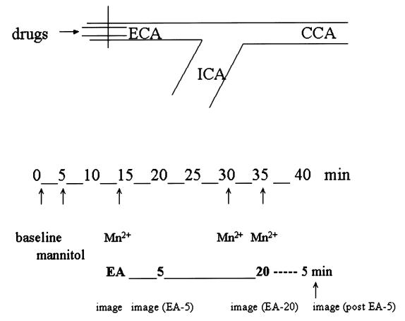
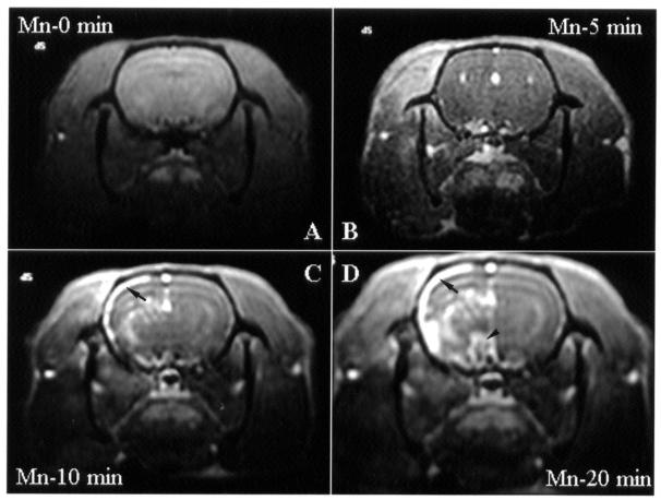
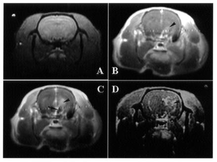
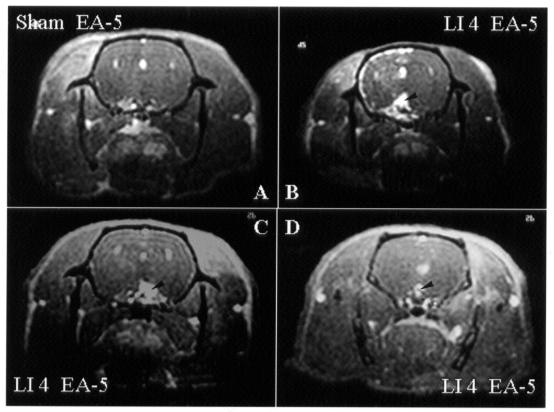
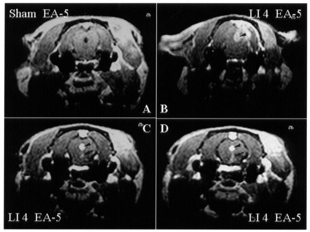
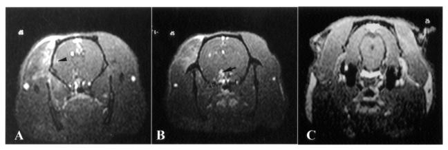
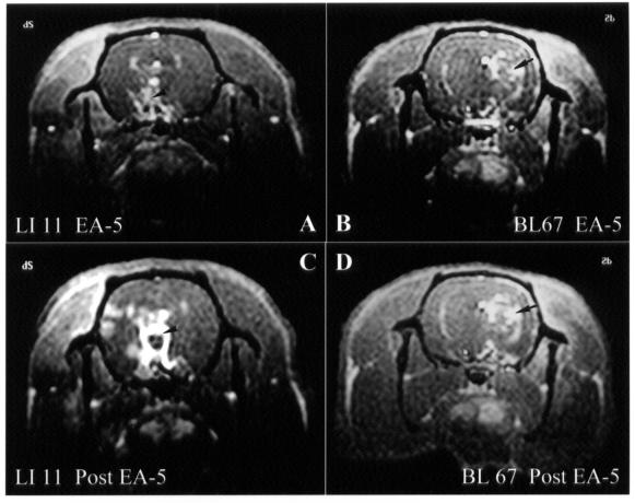
Acupuncture analgesia is an important issue in veterinary medicine. This study was designed to elucidate central modulation effects in response to electroacupuncture (EA) at different acupoints. Manganese-enhanced functional magnetic resonance imaging was performed in Sprague-Dawley rats after sham acupuncture, sham EA, or true EA at somatic acupoints. The acupoints were divided into 3 groups: group 1, analgesic acupoints commonly used for pain relief, such as Hegu (LI 4); group 2, nonanalgesic acupoints rarely used for analgesic effect such as Neiguan (PC 6); and group 3, acupoints occasionally used for analgesia, such as Zusanli (ST 36). Image acquisition was performed on a 1.5-T superconductive clinical scanner with a circular polarized extremity coil. The results showed that there was no neural activation caused by EA at a true acupoint with shallow needling and no electric current (sham acupuncture). When EA at a true acupoint was applied with true needling but no electric current (sham EA), there was only a slight increase in brain activity at the hypothalamus; when EA was applied at a true acupoint with true needling and an electric current (true EA), the primary response at the hypothalamus was enhanced. Also, there was a tendency for the early activation of pain-modulation areas to be prominent after EA at analgesic acupoints as compared with nonanalgesic acupoints. In conclusion, understanding the linkage between peripheral acupoint stimulation and central neural pathways provides not only an evidence-based approach for veterinary acupuncture but also a useful guide for clinical applications of acupuncture.
Figures
Similar articles
-
Neuronal specificity of needling acupoints at same meridian: a control functional magnetic resonance imaging study with electroacupuncture.Acupunct Electrother Res. 2007;32(3-4):179-93. Acupunct Electrother Res. 2007. PMID: 18494380
-
Electroacupuncture-induced neural activation detected by use of manganese-enhanced functional magnetic resonance imaging in rabbits.Am J Vet Res. 2001 Feb;62(2):178-82. doi: 10.2460/ajvr.2001.62.178. Am J Vet Res. 2001. PMID: 11212024
-
Electroacupuncture analgesia for surgery in cattle.Am J Chin Med. 2004;32(1):131-40. doi: 10.1142/S0192415X0400176X. Am J Chin Med. 2004. PMID: 15154292 Clinical Trial.
-
Neural mechanism underlying acupuncture analgesia.Prog Neurobiol. 2008 Aug;85(4):355-75. doi: 10.1016/j.pneurobio.2008.05.004. Epub 2008 Jun 5. Prog Neurobiol. 2008. PMID: 18582529 Review.
-
Acupuncture for Pain Management: Molecular Mechanisms of Action.Am J Chin Med. 2020;48(4):793-811. doi: 10.1142/S0192415X20500408. Epub 2020 May 15. Am J Chin Med. 2020. PMID: 32420752 Review.
Cited by
-
Analgesic Neural Circuits Are Activated by Electroacupuncture at Two Sets of Acupoints.Evid Based Complement Alternat Med. 2016;2016:3840202. doi: 10.1155/2016/3840202. Epub 2016 Jun 27. Evid Based Complement Alternat Med. 2016. PMID: 27429635 Free PMC article.
-
Mechanisms of acupuncture analgesia: effective therapy for musculoskeletal pain?Curr Rheumatol Rep. 2007 Dec;9(6):473-81. doi: 10.1007/s11926-007-0077-z. Curr Rheumatol Rep. 2007. PMID: 18177601 Review.
-
Effect of acupuncture 'dose' on modulation of the default mode network of the brain.Acupunct Med. 2016 Dec;34(6):425-432. doi: 10.1136/acupmed-2016-011071. Epub 2016 Oct 6. Acupunct Med. 2016. PMID: 27841974 Free PMC article. Clinical Trial.
-
How does moxibustion possibly work?Evid Based Complement Alternat Med. 2013;2013:198584. doi: 10.1155/2013/198584. Epub 2013 Mar 27. Evid Based Complement Alternat Med. 2013. PMID: 23606872 Free PMC article.
-
The mechanistic studies of acupuncture and moxibustion in Taiwan.Chin J Integr Med. 2011 Mar;17(3):177-86. doi: 10.1007/s11655-011-0664-8. Chin J Integr Med. 2011. PMID: 21359918 Review.
References
-
- Belliveau JW, Kennedy DN, McKinstry RC, et al. Functional mapping of the human visual cortex by magnetic resonance imaging. Science 1991;254:716–719. - PubMed
-
- Wiech K, Preissl H, Birbaumer N. Neural networks and pain processing. New insights from imaging techniques. Anaesthesist 2001;50:2–12. - PubMed
-
- Mathews KA. Nonsteroidal anti-inflammatory analgesics. Indications and contraindications for pain management in dogs and cats. Vet Clin North Am Small Anim Pract 2000;30:783–804. - PubMed
Publication types
MeSH terms
Substances
LinkOut - more resources
Full Text Sources
Medical