. 2003 Jun;71(6):3116-24.
doi: 10.1128/IAI.71.6.3116-3124.2003.

Relationship of plcR-regulated factors to Bacillus endophthalmitis virulence

Affiliations

Relationship of plcR-regulated factors to Bacillus endophthalmitis virulence

Michelle C Callegan et al. Infect Immun. 2003 Jun.

Abstract

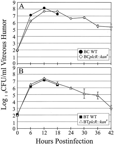
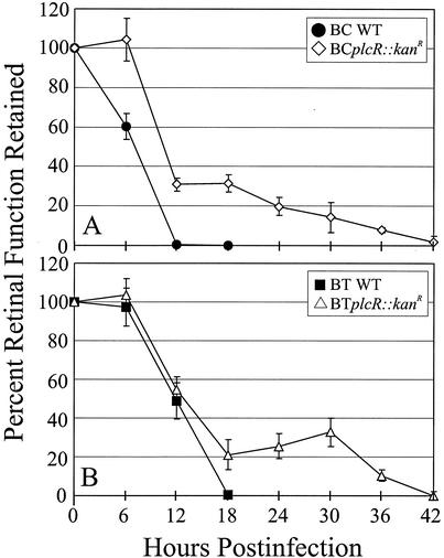
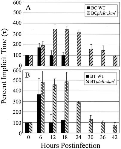
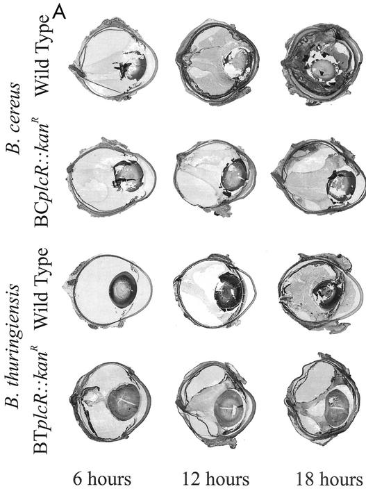
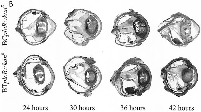
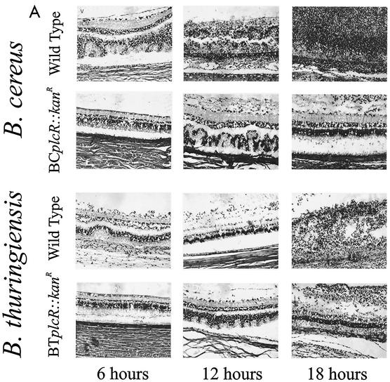
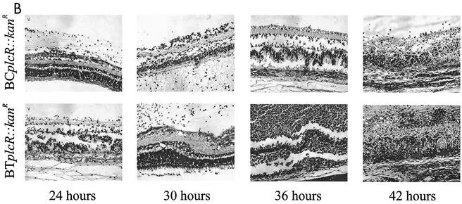
The explosive, destructive course of Bacillus endophthalmitis has been attributed to the production of toxins during infection. In this study we analyzed the contribution of toxins controlled by the global regulator plcR to the pathogenesis of experimental Bacillus endophthalmitis. Isogenic plcR-deficient mutants of Bacillus cereus and Bacillus thuringiensis were constructed by insertional inactivation of plcR by the kanamycin resistance cassette, aphA3. Rabbit eyes were injected intravitreally with approximately 100 CFU of wild-type B. cereus or B. thuringiensis or a plcR-deficient mutant. The evolution of endophthalmitis resulting from each plcR-deficient mutant was considerably slower than that caused by each wild-type strain. Retinal function was not eliminated until 42 h postinfection in rabbits with endophthalmitis caused by the plcR-deficient mutants, whereas wild-type infections resulted in a complete loss of retinal function within 18 h. The intraocular inflammatory cell influx and retinal destruction in plcR-deficient endophthalmitis approached the severity observed in wild-ype infections, but not until 36 h postinfection. Gross and histological examinations of eyes infected with plcR mutants demonstrated that the anterior and posterior segment changes were muted compared to the changes observed in eyes infected with the wild types. The loss of plcR-regulated factors significantly attenuated the severity of Bacillus endophthalmitis. The results therefore suggest that plcR may represent a target for which adjunct therapies could be designed for the prevention of blindness during Bacillus endophthalmitis.


Figures

FIG. 1.
FIG. 1.
Intraocular growth of B. cereus and B. thuringiensis wild-type strains and their plcR-deficient mutants. Approximately 100 CFU of each Bacillus strain was injected intravitreally. Wild-type Bacillus strains were quantified every 6 h for 18 h, while plcR-deficient strains were quantified every 6 h for 42 h of intraocular growth. The strains analyzed were B. cereus ATCC 14579 (BC WT) and its plcR-deficient mutant (BCplcR::kanR) (A) and B. thuringiensis BT407 (BT WT) and its plcR-deficient mutant (BTplcR::kanR) (B). All values are means ± SEM for four or more eyes per group.
FIG. 2.
FIG. 2.
Retinal function analysis of wild-type and plcR-deficient Bacillus endophthalmitis. ERG was performed every 6 h throughout the course of infection. Rapid decreases in b-wave amplitude were observed in eyes infected with wild-type Bacillus strains for 18 h, while gradual decreases were observed over a 42-h period in eyes infected with plcR mutant strains. All values are means ± SEM for four or more eyes per group. BC WT, B. cereus ATCC 14579; BCplcR::kanR, plcR-deficient mutant of B. cereus ATCC 14579; BT WT, B. thuringiensis BT407; BTplcR::kanR, plcR-deficient mutant of B. thuringiensis BT407.
FIG. 3.
FIG. 3.
Analysis of latent b-wave responses in wild-type and plcR-deficient Bacillus endophthalmitis. Implicit times (τ) from a-wave valleys to b-wave peaks of electroretinograms are shown. Increases in implicit times were observed at 6 h only for eyes infected with wild-type strains and were extended from 6 to 24 h for eyes infected with plcR-deficient strains. Increases in retinal response latencies may reflect changes in functional integrity associated with the rate of retinal response. All values are means ± SEM for four or more eyes per group. BC WT, B. cereus ATCC 14579; BCplcR::kanR, plcR-deficient mutant of B. cereus ATCC 14579; BT WT, B. thuringiensis BT407; BTplcR::kanR, plcR-deficient mutant of B. thuringiensis BT407.
FIG. 4.
FIG. 4.
Inflammatory cell influx into the anterior segment in wild-type and plcR-deficient Bacillus endophthalmitis. Anterior chamber paracentesis was performed to harvest aqueous humor, and inflammatory cells were quantified every 6 h throughout the course of infection. Rapid increases in the numbers of inflammatory cells were observed by 12 h, regardless of the infecting strain. The numbers of cells in plcR-infected eyes were similar to peak levels throughout 42 h of infection. All values are means ± SEM for four or more eyes per group. BC WT, B. cereus ATCC 14579; BCplcR::kanR, plcR-deficient mutant of B. cereus ATCC 14579; BT WT, B. thuringiensis BT407; BTplcR::kanR, plcR-deficient mutant of B. thuringiensis BT407.
FIG. 5.
FIG. 5.
(A) Whole-organ histological analysis of wild-type and plcR-deficient Bacillus endophthalmitis. In eyes infected with wild-type Bacillus, photoreceptor folding was observed in retinal sections by 12 h. By 18 h, severe inflammation was observed, and retinal layers were difficult to differentiate. Eyes infected with plcR-deficient strains showed mild inflammatory changes compared with eyes infected with wild-type strains at 12 and 18 h postinfection. All representative histological sections were stained with hematoxylin and eosin. Magnification, ×8.9. (B) Whole-organ histological analysis of extended plcR-deficient Bacillus endophthalmitis. Inflammatory changes were relatively mild throughout 30 h of infection. From 36 to 42 h, photoreceptor folding, random retinal detachments, and severe inflammation were observed. All representative histological sections were stained with hematoxylin and eosin. Magnification, ×7.5.
FIG. 5.
FIG. 5.
(A) Whole-organ histological analysis of wild-type and plcR-deficient Bacillus endophthalmitis. In eyes infected with wild-type Bacillus, photoreceptor folding was observed in retinal sections by 12 h. By 18 h, severe inflammation was observed, and retinal layers were difficult to differentiate. Eyes infected with plcR-deficient strains showed mild inflammatory changes compared with eyes infected with wild-type strains at 12 and 18 h postinfection. All representative histological sections were stained with hematoxylin and eosin. Magnification, ×8.9. (B) Whole-organ histological analysis of extended plcR-deficient Bacillus endophthalmitis. Inflammatory changes were relatively mild throughout 30 h of infection. From 36 to 42 h, photoreceptor folding, random retinal detachments, and severe inflammation were observed. All representative histological sections were stained with hematoxylin and eosin. Magnification, ×7.5.
FIG. 6.
FIG. 6.
(A) Retinal histological analysis of wild-type and plcR-deficient Bacillus endophthalmitis. Retinas from eyes infected with wild-type Bacillus were highly inflamed and partially detached, and the photoreceptor layers were indistinguishable by 18 h. Retinas from eyes infected with plcR-deficient Bacillus were mildly inflamed and essentially intact at 18 h postinfection. Abbreviations: V, vitreous; ILM, inner limiting membrane; GCL, ganglion cell layer; PC, photoreceptor cell layer; RPE, retinal pigment epithelium; CC, choriocapillaris. Magnification, ×168. (B) Retinal histological analysis of extended plcR-deficient Bacillus endophthalmitis. Retinas from eyes infected with plcR-deficient Bacillus were essentially intact, and there were small areas of photoreceptor layer folding by 30 h postinfection. Significant inflammation and photoreceptor layer folding were apparent by 36 h, and retinal layers were severely disrupted by 42 h postinfection. Magnification, ×128.
FIG. 6.
FIG. 6.
(A) Retinal histological analysis of wild-type and plcR-deficient Bacillus endophthalmitis. Retinas from eyes infected with wild-type Bacillus were highly inflamed and partially detached, and the photoreceptor layers were indistinguishable by 18 h. Retinas from eyes infected with plcR-deficient Bacillus were mildly inflamed and essentially intact at 18 h postinfection. Abbreviations: V, vitreous; ILM, inner limiting membrane; GCL, ganglion cell layer; PC, photoreceptor cell layer; RPE, retinal pigment epithelium; CC, choriocapillaris. Magnification, ×168. (B) Retinal histological analysis of extended plcR-deficient Bacillus endophthalmitis. Retinas from eyes infected with plcR-deficient Bacillus were essentially intact, and there were small areas of photoreceptor layer folding by 30 h postinfection. Significant inflammation and photoreceptor layer folding were apparent by 36 h, and retinal layers were severely disrupted by 42 h postinfection. Magnification, ×128.

References

    1. Abdelnour, A., S. Arvidson, T. Bremell, C. Ryden, and A. Tarkowski. 1993. The accessory gene regulator (agr) controls Staphylococcus aureus virulence in a murine arthritis model. Infect. Immun. 61:3879-3885. - PMC - PubMed
    1. Agaisse, H., M. Gominet, O. A. Okstad, A. B. Kolsto, and D. Lereclus. 1999. PlcR is a pleiotropic regulator of extracellular virulence factor gene expression in Bacillus thuringiensis. Mol. Microbiol. 32:1043-1053. - PubMed
    1. Arvidson, S., and K. Tegmark. 2001. Regulation of virulence determinants in Staphylococcus aureus. Int. J. Med. Microbiol. 291:159-170. - PubMed
    1. Association for Research in Vision and Ophthalmology. 2002, posing date. Statement for the use of animals in ophthalmic and visual research. [Online.] Association for Research in Vision and Ophthalmology, Rockville, Md. http://www.arvo.org/AboutARVO/animalst.htm.
    1. Block, F., and M. Schwarz. 1998. The b-wave of the electroretinogram as an index of retinal ischemia. Gen. Pharmacol. 30:281-287. - PubMed

Publication types

LinkOut - more resources