

The secreted aspartyl proteinases Sap1 and Sap2 cause tissue damage in an in vitro model of vaginal candidiasis based on reconstituted human vaginal epithelium
- PMID: 12761103
- PMCID: PMC155757
- DOI: 10.1128/IAI.71.6.3227-3234.2003
The secreted aspartyl proteinases Sap1 and Sap2 cause tissue damage in an in vitro model of vaginal candidiasis based on reconstituted human vaginal epithelium
Abstract
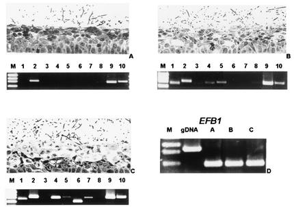
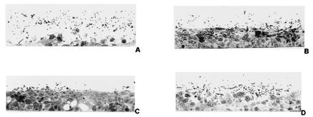
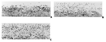
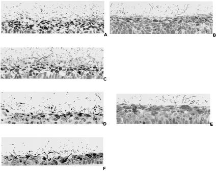
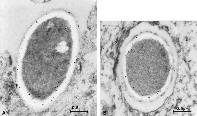
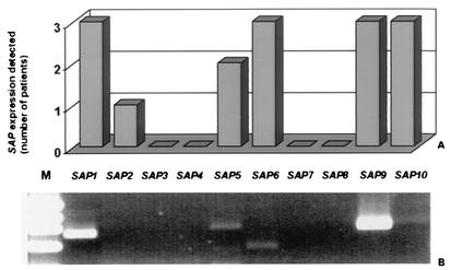
Secreted aspartyl proteinases (Saps) contribute to the ability of Candida albicans to cause mucosal and disseminated infections. A model of vaginal candidiasis based on reconstituted human vaginal epithelium (RHVE) was used to study the expression and role of these C. albicans proteinases during infection and tissue damage of vaginal epithelium. Colonization of the RHVE by C. albicans SC5314 did not cause any visible epithelial damage 6 h after inoculation, although expression of SAP2, SAP9, and SAP10 was detected by reverse transcriptase PCR. However, significant epithelial damage was observed after 12 h, concomitant with the additional expression of SAP1, SAP4, and SAP5. Additional transcripts of SAP6 and SAP7 were detected at a later stage of the artificial infection (24 h). Similar SAP expression profiles were observed in three samples isolated from human patients with vaginal candidiasis. In experimental infection, secretion of antigens Sap1 to Sap6 by C. albicans was confirmed at the ultrastructural level by using polyclonal antisera raised against Sap1 to Sap6. Addition of the aspartyl proteinase inhibitors pepstatin A and the human immunodeficiency virus proteinase inhibitors ritonavir and amprenavir strongly reduced the tissue damage of the vaginal epithelia by C. albicans cells. Furthermore, SAP null mutants lacking either SAP1 or SAP2 had a drastically reduced potential to cause tissue damage even though SAP3, SAP4, and SAP7 were up-regulated in these mutants. In contrast the vaginopathic potential of mutants lacking SAP3 or SAP4 to SAP6 was not reduced compared to wild-type cells. These data provide further evidence for a crucial role of Sap1 and Sap2 in C. albicans vaginal infections.
Figures
References
-
- Bain, J. M., C. Stubberfield, and N. A. Gow. 2001. Ura-status-dependent adhesion of Candida albicans mutants. FEMS Microbiol. Lett. 204:323-328. - PubMed
-
- Borg-von Zepelin, M., S. Beggah, K. Boggian, D. Sanglard, and M. Monod. 1998. The expression of the secreted aspartyl proteinases Sap4 to Sap6 from Candida albicans in murine macrophages. Mol. Microbiol. 28:543-554. - PubMed
-
- Calderone, R. A., and W. A. Fonzi. 2001. Virulence factors of Candida albicans. Trends Microbiol. 9:327-335. - PubMed
-
- Cassone, A., F. De Bernardis, F. Mondello, T. Ceddia, and L. Agatensi. 1987. Evidence for a correlation between proteinase secretion and vulvovaginal candidosis. J. Infect. Dis. 156:777-783. - PubMed
-
- De Bernardis, F., L. Agatensi, I. K. Ross, G. W. Emerson, R. Lorenzini, P. A. Sullivan, and A. Cassone. 1990. Evidence for a role for secreted aspartate proteinase of Candida albicans in vulvovaginal candidiasis. J. Infect. Dis. 161:1276-1283. - PubMed
Publication types
MeSH terms
Substances
LinkOut - more resources
Full Text Sources
Other Literature Sources
Molecular Biology Databases
Miscellaneous
