. 2003 Jun;71(6):3349-56.
doi: 10.1128/IAI.71.6.3349-3356.2003.

Structural and functional consequences of cleavage of human secretory and human serum immunoglobulin A1 by proteinases from Proteus mirabilis and Neisseria meningitidis

Affiliations

Structural and functional consequences of cleavage of human secretory and human serum immunoglobulin A1 by proteinases from Proteus mirabilis and Neisseria meningitidis

Adel Almogren et al. Infect Immun. 2003 Jun.

Abstract

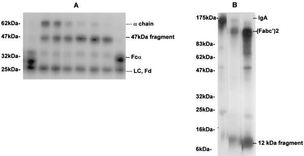
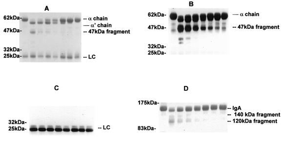
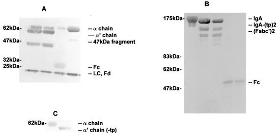
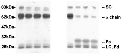
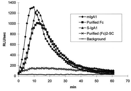
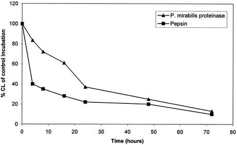
The cleavage of human serum monomeric immunoglobulin A1 (IgA1) and human secretory IgA1 (S-IgA1) by IgA1 proteinase of Neisseria meningitidis and cleavage by the proteinase from Proteus mirabilis have been compared. For serum IgA1, both proteinases cleaved only the alpha chain. N. meningitidis proteinase cleaved only in the hinge. P. mirabilis proteinase sequentially removed the tailpiece, the CH3 domain, and the CH2 domain. The cleavage of S-IgA1 by N. meningitidis proteinase occurred only in the hinge and was as rapid as that of serum IgA1. P. mirabilis proteinase predominantly cleaved the secretory component (SC) of S-IgA1. The SC of S-IgA1, whether cleaved or not, appeared to protect the alpha1 chain. Purified Fc fragment derived from the cleavage of serum IgA1 by N. meningitidis proteinase stimulated a respiratory burst in neutrophils through Fcalpha receptors, whereas the (Fcalpha1)(2)-SC fragment from digested S-IgA1 did not. The loss of the tailpiece from serum IgA1 treated with P. mirabilis proteinase had little effect, but the loss of the CH3 domain was concurrent with a rapid loss in the ability to bind to Fcalpha receptors. S-IgA1 treated with P. mirabilis proteinase under the same conditions retained the ability to bind to Fcalpha receptors. The results are consistent with the Fcalpha receptor binding site being at the CH2-CH3 interface. These data shed further light on the structure of S-IgA1 and indicate that the binding site for the Fcalpha receptor in S-IgA is protected by SC, thus prolonging its ability to activate phagocytic cells at the mucosal surface.


Figures

FIG. 1.
FIG. 1.
Time course of cleavage of human serum IgA1 by various proteinases and pepsin. (A) Cleavage of radiolabeled human serum IgA1 by proteinases from P. mirabilis, H. influenzae, and N. meningitidis. 125I-labeled serum IgA1 was incubated at 37°C with proteinases from H. influenzae for 4 h (lane 1); P. mirabilis for 4, 8, 16, 24, 32, or 48 h (lanes 4 to 9, respectively); or N. meningitidis for 4 h (lane 8). Products were analyzed by SDS-PAGE under reducing conditions and detected by autoradiography. LC, light chain. Positions of molecular mass markers are shown on the left. (B) Cleavage of radiolabeled human serum IgA1 by pepsin and P. mirabilis proteinase. 125I-labeled serum IgA1 was incubated at 37°C with buffer alone (lane 1), pepsin (lane 2), or P. mirabilis proteinase (lane 3) for 48 h. Products were analyzed by SDS-PAGE under nonreducing conditions and detected by autoradiography. Positions of molecular mass markers are shown on the left.
FIG. 2.
FIG. 2.
Cleavage of human serum IgA1 by P. mirabilis proteinase. Aliquots of 80 μg of serum IgA1 in 20 μl of buffer were incubated for 72 h at 37°C with 20 μl of buffer alone (lane 1) or at 37°C with 20 μl of purified P. mirabilis proteinase at 0.2 mg/ml (lane 2) or as serial doubling dilutions (lanes 3 to 8). The products (10 μg of IgA1) were analyzed on 5 to 20% gradient polyacrylamide gels run under reducing (A to C) or nonreducing (D) conditions. After the transfer of proteins to nitrocellulose membranes, the blots were stained with Ponceau S dye (A and D) or incubated with anti-α chain (B) or anti-light chain (LC) (C) alkaline phosphatase-conjugated antibodies and developed.
FIG. 3.
FIG. 3.
Cleavage of human serum IgA1 by purified proteinases from P. mirabilis and N. meningitidis. (A and B) Serum IgA1 was incubated at 37°C with buffer alone for 40 h (A, fourth lane; B, first lane), purified P. mirabilis proteinase for 24 and 40 h (A, first and second lanes; B, second and third lanes), or purified N. meningitidis proteinase for 8 h (A, third lane; B, fourth and fifth lanes). Samples were analyzed by SDS-PAGE under reducing (A) or nonreducing (B) conditions and gold stained. LC, light chain. (C) Immunoblot of recombinant IgA1 (lane1) and recombinant IgA1 with the tailpiece deleted (−tp) (lane 2) analyzed by SDS-PAGE under reducing conditions.
FIG. 4.
FIG. 4.
Cleavage of S-IgA1 by purified proteinases from P. mirabilis and N. meningitidis. S-IgA1 was incubated at 37°C with buffer alone for 40 h (first and fifth lanes); P. mirabilis proteinase for 8, 16, or 40 h (second through fourth lanes); or N. meningitidis proteinase for 2, 4, or 8 h (sixth to eighth lanes). Samples were analyzed by SDS-PAGE under reducing conditions and gold stained. LC, light chain.
FIG. 5.
FIG. 5.
Stimulation of neutrophil chemiluminescence by human serum monomeric IgA1 (mIgA1), S-IgA1, and the Fc fragments derived from them by cleavage with N. meningitidis proteinase. Wells of microtiter plates were coated in triplicate with human serum monomeric IgA1, S-IgA1, or the Fc fragments produced by their cleavage with N. meningitidis proteinase which had been purified by gel filtration with FPLC Superdex 200. Luminol-enhanced chemiluminescence was measured at 37°C for 60 min after the addition of purified neutrophils. The traces show the mean values for triplicate samples. RLU, relative light units.
FIG. 6.
FIG. 6.
Effect of digestion of human serum IgA1 with pepsin or P. mirabilis proteinase on its ability to stimulate neutrophil chemiluminescence. Serum IgA1 was left untreated or was cleaved at 37°C for up to 72 h with pepsin at pH 4.5 or with P. mirabilis proteinase at pH 8.0. The products were bound in duplicate to microtiter plates before the addition of purified neutrophils and Luminol. The mean chemiluminescence (CL) was measured and calculated as a percentage of the value for the undigested serum control. The P. mirabilis proteinase had cleaved the tailpiece from IgA1 by 5 h as well as the CH3 domain by 24 h.

References

    1. Boehm, M. K., J. M. Woof, M. A. Kerr, and S. J. Perkins. 1999. The Fab and Fc fragments of IgA1 exhibit a different arrangement from that in IgG: a study by X-ray and neutron solution scattering and homology modelling. J. Mol. Biol. 286:1421-1447. - PubMed
    1. Brooks, G. F., C. J. Lammel, M. S. Blake, B. Kusecek, and M. Achtman. 1992. Antibodies against IgA protease are stimulated both by clinical disease and asymptomatic carriage of serogroup A Neisseria meningitidis. J. Infect. Dis. 166:1316-1321. - PubMed
    1. Carayannopoulos, L., J. M. Hexam, and J. D. Capra. 1996. Localization of the binding site for the monocyte immunoglobulin (Ig) A-Fc receptor (CD89) to the domain boundary between Calpha2 and Calpha3 in human IgA1. J. Exp. Med. 183:1579-1586. - PMC - PubMed
    1. Devenyi, A. G., A. G. Plaut, F. J. Grundy, and A. Wright. 1993. Post-infectious human serum antibodies inhibit IgA1 proteinases by interaction with the cleavage site specificity determinant. Mol. Immunol. 30:1243-1248. - PubMed
    1. Gilbert, J. V., A. G. Plaut, B. Longmaid, and M. E. Lamm. 1983. Inhibition of microbial IgA proteases by human secretory IgA and serum. Mol. Immunol. 20:1039-1049. - PubMed

MeSH terms

LinkOut - more resources