

Host susceptibility to the attaching and effacing bacterial pathogen Citrobacter rodentium
- PMID: 12761129
- PMCID: PMC155702
- DOI: 10.1128/IAI.71.6.3443-3453.2003
Host susceptibility to the attaching and effacing bacterial pathogen Citrobacter rodentium
Abstract
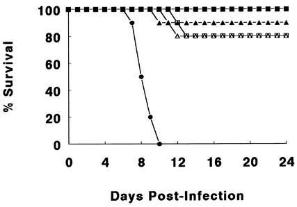
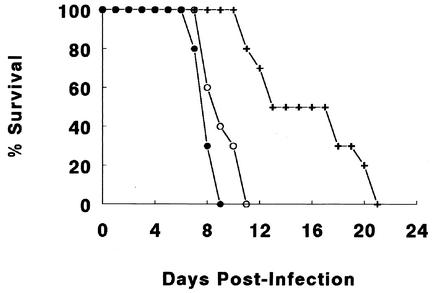
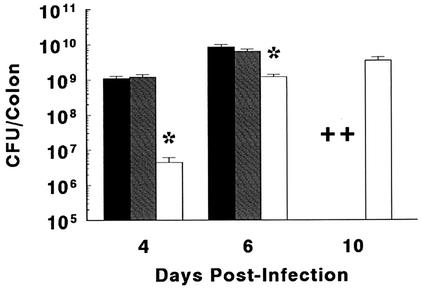
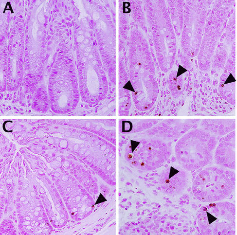
Many studies have shown that genetic susceptibility plays a key role in determining whether bacterial pathogens successfully infect and cause disease in potential hosts. Surprisingly, whether host genetics influence the pathogenesis of attaching and effacing (A/E) bacteria such as enteropathogenic and enterohemorrhagic Escherichia coli has not been examined. To address this issue, we infected various mouse strains with Citrobacter rodentium, a member of the A/E pathogen family. Of the strains tested, the lipopolysaccharide (LPS) nonresponder C3H/HeJ mouse strain experienced more rapid and extensive bacterial colonization than did other strains. Moreover, the high bacterial load in these mice was associated with accelerated crypt hyperplasia, mucosal ulceration, and bleeding, together with very high mortality rates. Interestingly, the basis for the increased susceptibility was not due to LPS hyporesponsiveness, as the genetically related but LPS-responsive C3H/HeOuJ and C3H/HeN mouse strains were also susceptible to infection. Analysis of the intestinal pathology in these susceptible strains revealed significant crypt epithelial cell apoptosis (terminal deoxynucleotidyltransferase-mediated dUTP-biotin nick end label staining) as well as bacterial translocation to the mesenteric lymph nodes. Further studies with infection of SCID (T- and B-lymphocyte-deficient) C3H/HeJ mice demonstrated that loss of lymphocytes had no effect on bacterial numbers but did reduce crypt cell apoptosis and delayed mortality. These studies thus identify the adaptive immune system, crypt cell apoptosis, and bacterial translocation but not LPS responsiveness as contributing to the tissue pathology and mortality seen during C. rodentium infection of highly susceptible mouse strains. Determining the basis for these strains' susceptibility to intestinal colonization by an A/E pathogen will be the focus of future studies.
Figures

Similar articles
-
Central role for B lymphocytes and CD4+ T cells in immunity to infection by the attaching and effacing pathogen Citrobacter rodentium.Infect Immun. 2003 Sep;71(9):5077-86. doi: 10.1128/IAI.71.9.5077-5086.2003. Infect Immun. 2003. PMID: 12933850 Free PMC article.
-
Impaired resistance and enhanced pathology during infection with a noninvasive, attaching-effacing enteric bacterial pathogen, Citrobacter rodentium, in mice lacking IL-12 or IFN-gamma.J Immunol. 2002 Feb 15;168(4):1804-12. doi: 10.4049/jimmunol.168.4.1804. J Immunol. 2002. PMID: 11823513
-
Toll-like receptor 4 contributes to colitis development but not to host defense during Citrobacter rodentium infection in mice.Infect Immun. 2006 May;74(5):2522-36. doi: 10.1128/IAI.74.5.2522-2536.2006. Infect Immun. 2006. PMID: 16622187 Free PMC article.
-
Molecular pathogenesis of Citrobacter rodentium and transmissible murine colonic hyperplasia.Microbes Infect. 2001 Apr;3(4):333-40. doi: 10.1016/s1286-4579(01)01387-9. Microbes Infect. 2001. PMID: 11334751 Review.
-
Overview of the Effect of Citrobacter rodentium Infection on Host Metabolism and the Microbiota.Methods Mol Biol. 2021;2291:399-418. doi: 10.1007/978-1-0716-1339-9_20. Methods Mol Biol. 2021. PMID: 33704766 Review.
Cited by
-
Innate Lymphoid Cells Control Early Colonization Resistance against Intestinal Pathogens through ID2-Dependent Regulation of the Microbiota.Immunity. 2015 Apr 21;42(4):731-43. doi: 10.1016/j.immuni.2015.03.012. Immunity. 2015. PMID: 25902484 Free PMC article.
-
Comparative Genomic Analysis of Citrobacter and Key Genes Essential for the Pathogenicity of Citrobacter koseri.Front Microbiol. 2019 Dec 6;10:2774. doi: 10.3389/fmicb.2019.02774. eCollection 2019. Front Microbiol. 2019. PMID: 31866966 Free PMC article.
-
Immunization of mice with Lactobacillus casei expressing a beta-intimin fragment reduces intestinal colonization by Citrobacter rodentium.Clin Vaccine Immunol. 2011 Nov;18(11):1823-33. doi: 10.1128/CVI.05262-11. Epub 2011 Sep 7. Clin Vaccine Immunol. 2011. PMID: 21900533 Free PMC article.
-
The pathogenicity of an enteric Citrobacter rodentium Infection is enhanced by deficiencies in the antioxidants selenium and vitamin E.Infect Immun. 2011 Apr;79(4):1471-8. doi: 10.1128/IAI.01017-10. Epub 2011 Jan 18. Infect Immun. 2011. PMID: 21245271 Free PMC article.
-
Putting the microbiota to work: Epigenetic effects of early life antibiotic treatment are associated with immune-related pathways and reduced epithelial necrosis following Salmonella Typhimurium challenge in vitro.PLoS One. 2020 Apr 27;15(4):e0231942. doi: 10.1371/journal.pone.0231942. eCollection 2020. PLoS One. 2020. PMID: 32339193 Free PMC article.
References
-
- Asfaha, S., W. K. MacNaughton, C. B. Appleyard, K. Chadee, and J. L. Wallace. 2001. Persistent epithelial dysfunction and bacterial translocation after resolution of intestinal inflammation. Am. J. Physiol. Gastrointest. Liver Physiol. 281:G635-G644. - PubMed
-
- Barthold, S. W., G. L. Coleman, P. N. Bhatt, G. W. Osbaldiston, and A. M. Jonas. 1976. The etiology of transmissible murine colonic hyperplasia. Lab. Anim. Sci. 26:889-894. - PubMed
-
- Barthold, S. W., G. W. Osbaldiston, and A. M. Jonas. 1977. Dietary, bacterial, and host genetic interactions in the pathogenesis of transmissible murine colonic hyperplasia. Lab. Anim. Sci. 27:938-945. - PubMed
Publication types
MeSH terms
Substances
LinkOut - more resources
Full Text Sources
Other Literature Sources
