

Neural correlates of outcome after stroke: a cross-sectional fMRI study
- PMID: 12764063
- PMCID: PMC3717456
- DOI: 10.1093/brain/awg145
Neural correlates of outcome after stroke: a cross-sectional fMRI study
Abstract
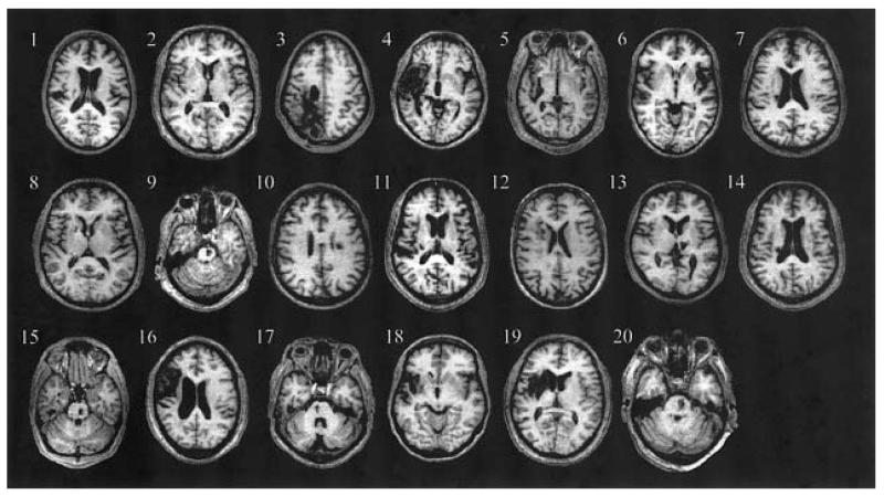
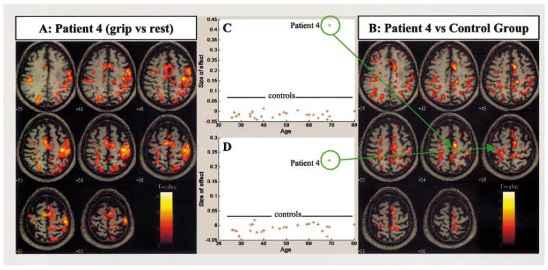
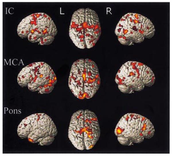
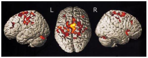
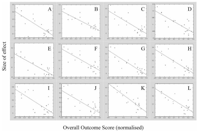
Recovery of motor function after stroke may occur over weeks or months and is often attributed to neuronal reorganization. Functional imaging studies investigating patients who have made a good recovery after stroke have suggested that recruitment of other motor-related networks underlies this recovery. However, patients with less complete recovery have rarely been studied, or else the degree of recovery has not been taken into account. We set out to investigate the relationship between the degree of recovery after stroke and the pattern of recruitment of brain regions during a motor task as measured using functional MRI. We recruited 20 patients who were at least 3 months after their first ever stroke, and 26 right-handed age-matched control subjects. None of our patients had infarcts involving the hand region of the primary motor cortex. All subjects were scanned whilst performing an isometric, dynamic visually paced handgrip task. The degree of functional recovery of each patient was assessed using a battery of outcome measures. Single-patient versus control group analysis revealed that patients with poor recovery were more likely to recruit a number of motor-related brain regions over and above those seen in the control group during the motor task, whereas patients with more complete recovery were more likely to have 'normal' task-related brain activation. Across the whole patient group and across stroke subtypes, we were able to demonstrate a negative correlation between outcome and the degree of task-related activation in regions such as the supplementary motor area, cingulate motor areas, premotor cortex, posterior parietal cortex, and cerebellum. This negative correlation was also seen in parts of both contralateral and ipsilateral primary motor cortex. These results further our understanding of the recovery process by demonstrating for the first time a clear relationship between task-related activation of the motor system and outcome after stroke.
Figures
References
-
- Allison JD, Meador KJ, Loring DW, Figueroa RE, Wright JC. Functional MRI cerebral activation and deactivation during finger movement. Neurology. 2000;54:135–42. - PubMed
-
- Andersson JL, Hutton C, Ashburner J, Turner R, Friston K. Modeling geometric deformations in EPI time series. Neuroimage. 2001;13:903–19. - PubMed
-
- Brett M, Leff AP, Rorden C, Ashburner J. Spatial normalization of brain images with focal lesions using cost function masking. Neuroimage. 2001;14:486–500. - PubMed
-
- Brinkman J, Kuypers HG. Cerebral control of contralateral and ipsilateral arm, hand and finger movements in the split-brain rhesus monkey. Brain. 1973;96:653–74. - PubMed
-
- Buchel C, Holmes AP, Rees G, Friston KJ. Characterizing stimulus-response functions using nonlinear regressors in parametric fMRI experiments. Neuroimage. 1998;8:140–8. - PubMed
Publication types
MeSH terms
Grants and funding
LinkOut - more resources
Full Text Sources
Medical
