

Review
doi: 10.1523/JNEUROSCI.23-10-03963.2003.
The underpinnings of the BOLD functional magnetic resonance imaging signal
Affiliations
- PMID: 12764080
- PMCID: PMC6741096
- DOI: 10.1523/JNEUROSCI.23-10-03963.2003
Item in Clipboard
Review
The underpinnings of the BOLD functional magnetic resonance imaging signal
J Neurosci.
.
No abstract available
Figures
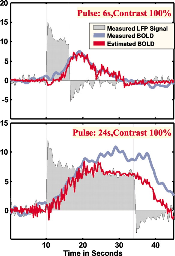
Measured responses as well as the measured and estimated BOLD response. Residual analysis showed increased error for longer pulse duration. Visual inspection of the data from 24-sec-long stimulus presentation revealed greatly increased residuals after the initial ramp of the BOLD response, suggesting lack of time invariance and the existence of nonlinearities not captured by the Wiener–Kernel analysis applied here to the data.
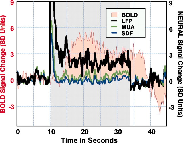
Simultaneous neural and hemodynamic recordings from a cortical site showing transient neural response. Note that both single-and multiple-unit responses adapt a couple of seconds after stimulus onset, with LFP remaining the only signal correlated with the BOLD response. The spike density function (SDF) reflects the instantaneous firing rate of a small population of neurons isolated during the analysis by using standard mathematical methods.
References
-
- Aika Y, Ren JQ, Kosaka K, Kosaka T ( 1994) Quantitative analysis of GABA-like-immunoreactive and parvalbumin-containing neurons in the CA1 region of the rat hippocampus using a stereological method, the dissector. Exp Brain Res 99: 267–276. - PubMed
-
- Ajmone-Marsan C ( 1965) Electrical activity of the brain: slow waves and neuronal activity. Israel J Med Sci 1: 104–117. - PubMed
-
- Arezzo J, Legatt AD, Vaughan HGJ ( 1979) Topography and intracranial sources of somatosensory evoked potentials in the monkey. I. Early components. Electroencephalogr Clin Neurophysiol 46: 155–172. - PubMed
Publication types
MeSH terms
Substances
LinkOut - more resources
Full Text Sources
Medical