

Review
doi: 10.1523/JNEUROSCI.23-10-03981.2003.
Neuroimaging weighs in: humans meet macaques in "primate" visual cortex
Affiliations
- PMID: 12764082
- PMCID: PMC6741079
- DOI: 10.1523/JNEUROSCI.23-10-03981.2003
Item in Clipboard
Review
Neuroimaging weighs in: humans meet macaques in "primate" visual cortex
J Neurosci.
.
No abstract available
Figures
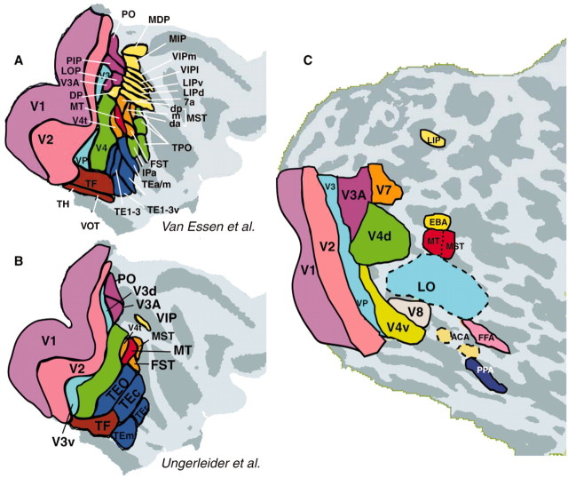
Maps of reported areas in primate visual cortex. Maps are shown on the flattened cortical surface from right hemisphere (light gray, gyri; dark gray, sulci). A shows areas in macaque reported by Van Essen and colleagues, and B shows the macaque areas reported by Ungerleider and collaborators (adapted from Van Essen et al., 2001). C shows areas in human visual cortex, as described in the text. Consensus is highest in lower-tier (generally, left-most) areas; such areas tend to be evolutionarily more conserved, and the retinotopy is more easily resolved.
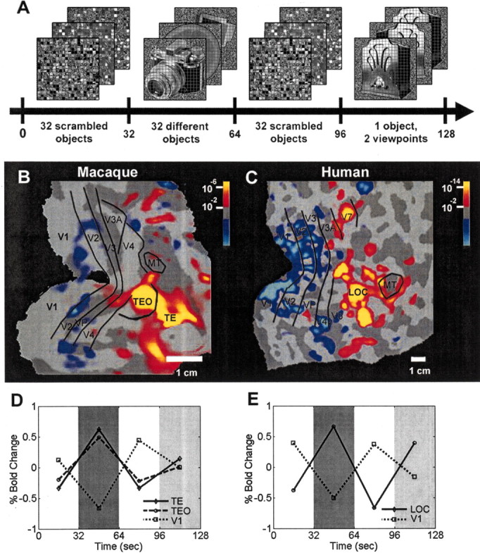
Object-selective (LOc) activation in visual cortex of macaques and humans. In both species, fMRI (BOLD) data were acquired from awake subjects, who fixated the center of a common stimulus set. A shows those stimuli, presented in block design in an a–b–a–c sequence (a, 32 grid-scrambled objects; b, 32 different objects; c, one object, presented in two different views). B (macaque) and C (human) reveal regions activated more by objects than scrambled objects (red-orange) and the reverse (blue-cyan). D (macaque) and E (human) show corresponding fMRI levels in selected visual areas. The human region activated more by objects (C) has been named LOc; it corresponds primarily to higher-tier cortical areas TEO and TE in macaque (B). In both species, lower-tier retinotopic areas (e.g., V1, V2, V3) responded better to the control images (scrambled objects), making the reversal in higher-tier areas even more significant. In human LOc and macaque TEO/TE, there was a reduced response to presentations of the single object (condition c, dark gray) compared with the multiple objects (condition b, light gray). Thus macaque shows fMRI-based adaptation in inferotemporal cortex, similar to that in humans. Bold, Blood oxygen level dependent.
References
-
- Aguirre GK, Singh R, D'Esposito M ( 1999) Stimulus inversion and the responses of face and object-sensitive cortical areas. NeuroReport 10: 189–194. - PubMed
-
- Allison T, Puce A, Spencer DD, McCarthy G ( 1999) Electrophysiological studies of human face perception. I: Potentials generated in occipitotemporal cortex by face and non-face stimuli. Cereb Cortex 9: 415–430. - PubMed
-
- Allman JM, Kaas JH, Lane RH ( 1973) The middle temporal visual area (MT) in the bushbaby, Galago senegalensis Brain Res 57: 197–202. - PubMed
-
- Backus BT, Fleet DJ, Parker AJ, Heeger DJ ( 2001) Human cortical activity correlates with stereoscopic depth perception. J Neurophysiol 86: 2054–2068. - PubMed
-
- Baker JF, Petersen SE, Newsome WT, Allman JM ( 1981) Visual response properties of neurons in four extrastriate visual areas of the owl monkey (Aotus trivirgatus): a quantitative comparison of medial, dorsomedial, dorsolateral, and middle temporal areas. J Neurophysiol 45: 397–416. - PubMed
Publication types
MeSH terms
LinkOut - more resources
Full Text Sources
Medical