. 2003 May 15;23(10):4034-43.
doi: 10.1523/JNEUROSCI.23-10-04034.2003.

Exploitation of astrocytes by glioma cells to facilitate invasiveness: a mechanism involving matrix metalloproteinase-2 and the urokinase-type plasminogen activator-plasmin cascade

Affiliations

Exploitation of astrocytes by glioma cells to facilitate invasiveness: a mechanism involving matrix metalloproteinase-2 and the urokinase-type plasminogen activator-plasmin cascade

Duc M Le et al. J Neurosci. .

Abstract

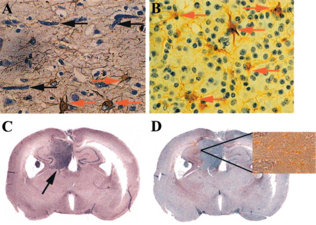
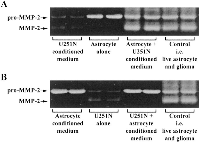
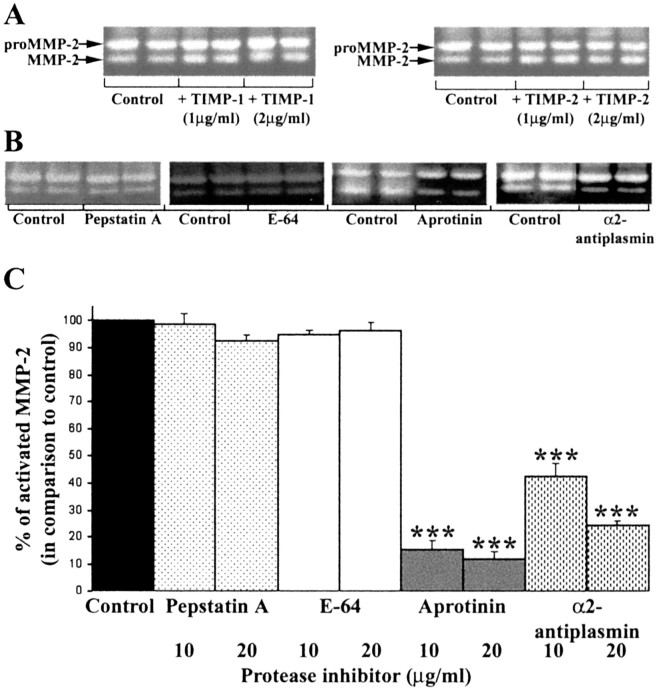
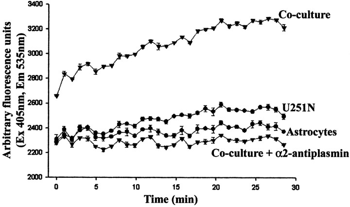
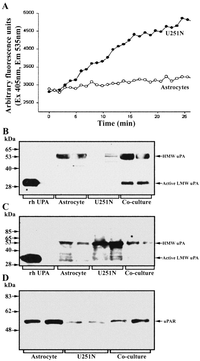
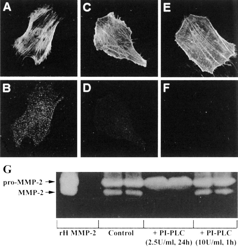
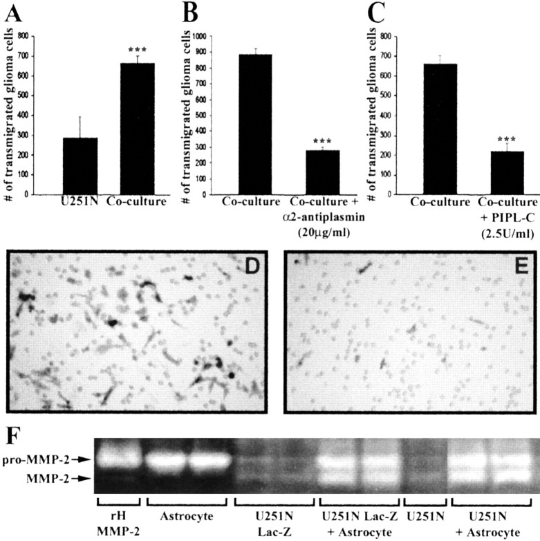
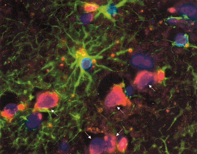
The presence of reactive astrocytes around glioma cells in the CNS suggests the possibility that these two cell types could be interacting. We addressed whether glioma cells use the astrocyte environment to modulate matrix metalloproteinase-2 (MMP-2), a proteolytic enzyme implicated in the invasiveness of glioma cells. We found that astrocytes in culture produce significant amounts of the pro-form of MMP-2 but undetectable levels of active MMP-2. However, after coculture with the U251N glioma line, astrocyte pro-MMP-2 was converted to the active form. The mechanism of pro-MMP-2 activation in glioma-astrocyte coculture was investigated and was found to involve the urokinase-type plasminogen activator (uPA)-plasmin cascade whereby uPA bound to uPA receptor (uPAR), leading to the conversion of plasminogen to plasmin. The latter cleaved pro-MMP-2 to generate its active form. Furthermore, key components (i.e., uPAR, uPA, and pro-MMP-2) were contributed principally by astrocytes, whereas the U251N glioma cells provided plasminogen. In correspondence with this biochemical cascade, the transmigration of U251N cells through Boyden invasion chambers coated with an extracellular matrix barrier was increased significantly in the presence of astrocytes, and this was inhibited by agents that disrupted the uPA-plasmin cascade. Finally, using resected human glioblastoma specimens, we found that tumor cells, but not astrocytes, expressed plasminogen in situ. We conclude that glioma cells exploit their astrocyte environment to activate MMP-2 and that this leads to the increased invasiveness of glioma cells.


Figures

Figure 1.
Figure 1.
Reactive astrocytes are found in proximity to glioma cellsin situ. Immunoreactivity for GFAP (brown cells) reveals reactive astrocytes in the proximity of glioma cells; samples are counterstained with hematoxylin. A, This section from a patient with malignant astrocytoma shows elongated glioma cells (black arrows) that have infiltrated into the cortex (at least 3 cm away) from the main tumor mass in a subcortical region. Reactive astrocytes are depicted with red arrows. B, Astrocytes (red arrows) are seen reacting to oligodendroglioma cells within the mass of the tumor. C, Significant tumor growth (indicated by the black arrow) in the hippocampus is observed in the brains of SCID mice 25d after implantation of U87 glioma cells, as revealed with hematoxylin and eosin staining. An adjacent section in D shows increased levels of GFAP immunoreactivity (brown speckles in the coronal section) in the tumor–brain interface. The inset in D is a high-power view of the indicated area to better display the reactive astrocytes. Similar results were observed for implanted U251N cells (data not shown).
Figure 2.
Figure 2.
Activation of pro-MMP-2 in cocultures of astrocytes and U251N glioma cells. A Gelatin zymogram of pro- and active MMP-2 is displayed. Recombinant human (rH) MMP-2 was used as a standard for both pro- and active MMP-2, which were ∼72 and 65 kDa, respectively. Although the conditioned medium of astrocytes contained high levels of pro-MMP-2 and negligible active MMP-2, U251N glioma cells secreted very low but detectable levels of both forms. In the coculture of astrocytes with U251N cells, pro-MMP-2 remained high; significantly, a substantial amount of active MMP-2 was generated. This result has been replicated in >20 experiments in which each test condition was always performed in duplicate (duplicate lanes per condition are displayed for this and other zymograms). B, The amount of active MMP-2 present in the conditioned medium of cells is documented by degradation of fluorescent substrate. In correspondence with the zymography data, increased MMP-2 activity was generated in U251N glioma–astrocyte coculture. The media histogram refers to feeding medium that had not been exposed to cells. Values shown are the mean±SEM of triplicate samples from a single experiment. This result has been reproduced in two other experiments; ***p < 0.001 compared against U251N glioma, one-way ANOVA with Bonferroni post hoc test. C, Shown is the GFAP status of cells, whereby the U251N cells (left panel) were found to contain GFAP intermediate filament protein (green stain) as do the fetal astrocytes (right panel); in the middle panel the primary anti-GFAP antibody has been omitted from the staining of U251N cells. All cells were counterlabeled with Hoechst dye to illuminate their nuclei. The presence of GFAP immunoreactivity in the U251 line used here indicates that the results of this manuscript are not attributable to the loss of GFAP expression, which can confer to glioma cells a more invasive phenotype (Rutka and Smith, 1993).
Figure 3.
Figure 3.
Pro-MMP-2 activation occurs in the presence of live astrocytes and glioma-soluble factors. The coculturing of live astrocytes and live U251N cells (Control) resulted in high levels of active MMP-2 (last 2 lanes of A, B). However, activation of MMP-2 required the presence of live astrocytes if glioma-conditioned medium were present (A). Activation of MMP-2 did not occur when live glioma cells were incubated with astrocyte-conditioned medium (B). This result was replicated in three separate experiments. For comparison, duplicate lanes of conditioned media from U251N or astrocytes or of cells alone are displayed also.
Figure 4.
Figure 4.
The use of protease inhibitors implicates plasmin in the activation of pro-MMP-2 in glioma–astrocyte cocultures. A, The addition of TIMP-1 and -2 did not affect the activation of MMP-2 in glioma–astrocyte cocultures, thereby excluding the participation of MMPs. Amixture of TIMP-1 (2 μg/ml) and TIMP-2 (2 μg/ml) was also ineffective at preventing pro-MMP-2 activation (data not shown). B, Shown are representative gelatin zymograms of conditioned media collected after 24 hr for U251N–astrocyte cocultures with (20 μg/ml) or without (Control) protease inhibitors; each condition was assessed in duplicate. Pepstatin A and E-64 did not block pro-MMP-2 activation, whereas aprotinin, an inhibitor of serine proteinases, and α2-antiplasmin (an inhibitor of the serine proteinase plasmin) attenuated active MMP-2 levels. These results were replicated in five experiments. C, The level of active MMP-2 generated in five cocultures of U251N–astrocyte was normalized to control cultures; then the level of active MMP-2 in treated cultures was expressed as a percentage of this normalized control. Values are the mean ± SEM; ***p < 0.001 compared with control, one-way ANOVA with Bonferroni post hoc comparisons. Thus the activation of MMP-2 is attenuated by aprotinin and α2-antiplasmin, implicating plasmin as an intermediary in the activation of MMP-2 in glioma–astrocyte cocultures.
Figure 5.
Figure 5.
Plasmin activity is high in U251N–astrocyte cocultures. Plasmin activity was assessed with a fluorochrome-conjugated plasmin substrate, AFC-80, and the increase in fluorescence over time was monitored. Conditioned media of astrocytes or U251N cells in isolation had low levels of plasmin activity, but, when the cells were cocultured, high plasmin activity was obtained. That the enzymatic activity was attributable to plasmin was shown by the inhibition of activity in cocultures to which α2-antiplasmin (10 μg/ml) was supplemented. This experiment was replicated three times with similar results. Note that the fluorescent intensity graph for the coculture starts from a higher point than for the other conditions that were examined because the degradation of the fluorogenic substrate occurs instantaneously in the coculture-conditioned medium, and we could not catch the initial plasmin level in time.
Figure 6.
Figure 6.
Contribution of components of the uPAR–plasmin cascade by astrocytes or glioma cells. A, Plasminogen content was determined by the use of the fluorochrome-conjugated plasmin substrate AFC-80; here, 10 μM uPA was applied to conditioned media to convert plasminogen to plasmin, the activity of which was monitored. Note that the U251N glioma cells (open circles), rather than astrocytes (filled circles), were the principal sources of plasminogen. This result was replicated three times. B, C, Western blots of cell lysates or conditioned media, respectively, of uPA expression by astrocytes and U251N cells. The cell lysate analyses (B) show high uPA levels for astrocytes, but not for U251N cells, whereas the converse is true for conditioned media (C). D, Western blot for uPAR that documents higher expression in astrocytes rather than in glioma cells.
Figure 7.
Figure 7.
Detection of uPAR on astrocytes by immunofluorescence. Astrocytes have high expression of uPAR (B), whereas U251N cells have very low levels (D). Astrocytes (A) and glioma cells (C) were counterstained with phalloidin, which binds to filamentous actin, to delineate the boundary of each cell type. The specificity of the uPAR antibody was tested with an uPAR peptide (Santa Cruz Biotechnology, Santa Cruz, CA); when the uPAR antibody was preincubated with the uPAR peptide, subsequent staining of astrocytes with this antibody mixture was negative (data not shown). E, F, Astrocytes were treated with PI-PLC (2.5 U/ml) for 24 hr and then immunostained for uPAR. After PI-PLC treatment the astrocytes (E; phalloidin staining) were no longer immunoreactive for uPAR (F). Finally, continuous 2.5 U/ml PI-PLC for 24 hr (but not 10 U/ml PI-PLC for 1 hr, followed by 23 hr of recovery) abrogated the activation of MMP-2 in glioma–astrocyte interaction (G).
Figure 8.
Figure 8.
Inhibition of plasmin or the removal of uPAR by PI-PLC decreases transmigration of U251N cells in invasion assays. Lac-Z U251N cells (25,000 cells) cocultured with astrocytes (10,000 cells) resulted in a high number of glioma cells transmigrating through the Matrigel-coated Boyden invasion chambers by 68 hr (A). The addition to the coculture of α2-antiplasmin (B) or PI-PLC (C; with PI-PLC replenished at 24 and 48 hr) significantly decreased the number of U251N cells invading through the chamber. This experiment was replicated three times. Values are the mean ± SEM of triplicate samples; Student's t test, ***p < 0.001. D, E, Representative micrographs of U251N cells, identified after staining for β-galactosidase, that have invaded across Matrigel in a control coculture (D) or in a coculture treated with inhibitors (E). The holes in D and E are the 8 μm pores present in the filter that supports the Matrigel barrier. F, Control experiment to indicate that the Lac-Z transfectants (U251N Lac-Z) indeed are behaving in a similar manner to the parent line (U251N) with respect to the activation of MMP-2 in glioma–astrocytes cocultures.
Figure 9.
Figure 9.
Plasminogen is expressed by glioma cells, but not by astrocytes, in vivo. A human glioblastoma brain section was immunolabeled for GFAP (green) to identify astrocytes that were in close proximity to tumor cells that were GFAP-negative; note that we selected specifically a glioblastoma sample that was GFAP-negative for glioma cells to allow for differentiation from astrocytes. Tumor cells were identified further by the size of their nuclei, which were larger than those of glia, and by the use of phase-contrast microscopy, whereby they could be distinguished from neurons with apical dendrites and a pyramidal morphology. Note that plasminogen (red) is expressed by the glioma cells (arrows). All nuclei were labeled by Hoechst dye (blue). We counted the proportions of glioma or astrocytes at the leading edge of tumor that were positive for plasminogen expression; three high-powered fields were sampled. Although no reactive astrocytes were scored positive, 19 of 23 (83%) convincingly identified glioma cells expressed plasminogen. Original magnification, 1000×.

Similar articles

Cited by

References

    1. Andreasen PA, Egelund R, Petersen HH ( 2000) The plasminogen activation system in tumor growth, invasion, and metastasis. Cell Mol Life Sci 57: 25–40. - PMC - PubMed
    1. Apodaca G, Rutka JT, Bouhana K, Berens ME, Giblin JR, Rosenblum ML, McKerrow JH, Banda MJ ( 1990) Expression of metalloproteinases and metalloproteinase inhibitors by fetal astrocytes and glioma cells. Cancer Res 50: 2322–2329. - PubMed
    1. Beliveau R, Delbecchi L, Beaulieu E, Mousseau N, Kachra Z, Berthelet F, Moumdjian R, Del Maestro R ( 1999) Expression of matrix metalloproteinases and their inhibitors in human brain tumors. Ann NY Acad Sci 886: 236–239. - PubMed
    1. Bello L, Lucini V, Carrabba G, Giussani C, Machluf M, Pluderi M, Nikas D, Zhang J, Tomei G, Villani RM, Carroll RS, Bikfalvi A, Black PM ( 2001) Simultaneous inhibition of glioma angiogenesis, cell proliferation, and invasion by a naturally occurring fragment of human metalloproteinase-2. Cancer Res 61: 8730–8736. - PubMed
    1. Benbow U, Schoenermark MP, Mitchell TI, Rutter JL, Shimokawa K, Nagase H, Brinckerhoff CE ( 1999) A novel host/tumor cell interaction activates matrix metalloproteinase-1 and mediates invasion through type I collagen. J Biol Chem 274: 25371–25378. - PubMed

Publication types

MeSH terms

LinkOut - more resources