

Endocytosis and vesicle recycling at a ribbon synapse
- PMID: 12764096
- PMCID: PMC6741077
- DOI: 10.1523/JNEUROSCI.23-10-04092.2003
Endocytosis and vesicle recycling at a ribbon synapse
Abstract
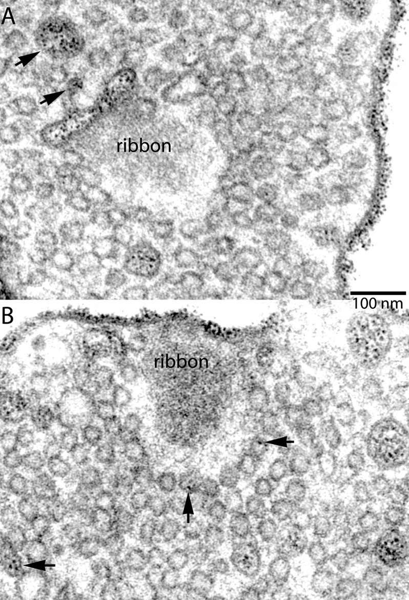
At ribbon synapses, where exocytosis is regulated by graded depolarization, vesicles can fuse very rapidly with the plasma membrane (complete discharge of the releasable pool in approximately 200 msec). Vesicles are also retrieved very rapidly (time constant of approximately 1 sec), leading us to wonder whether their retrieval uses an unusual mechanism. To study this, we exposed isolated bipolar neurons from goldfish retina to cationized ferritin. This electron-dense marker uniformly decorated the cell membrane and was carried into the cell during membrane retrieval. Endocytosis was activity-dependent and restricted to the synaptic terminal. The labeling pattern was consistent with direct retrieval from the plasma membrane of large, uncoated endosomes 60-200 nm in diameter. Even after extensive synaptic activity lasting several minutes, most of the ferritin remained in large endosomes and was present in only approximately 10% of the small vesicles that constitute the reserve pool. By contrast, after brief stimulation at a conventional terminal, ferritin did not reside in endosomes but was present in approximately 63% of the small vesicles. We suggest that the bipolar ribbon synapse sustains its rapid exocytosis by retrieving membrane in larger "bites" than the clathrin-dependent mechanism thought to dominate at conventional synapses. The resulting large endosomes bud off small vesicles, which reenter the reserve pool and finally the releasable pool.
Figures


Similar articles
-
The kinetics of exocytosis and endocytosis in the synaptic terminal of goldfish retinal bipolar cells.J Physiol. 1999 Feb 15;515 ( Pt 1)(Pt 1):181-202. doi: 10.1111/j.1469-7793.1999.181ad.x. J Physiol. 1999. PMID: 9925888 Free PMC article.
-
Distribution of proteins associated with synaptic vesicle endocytosis in the mouse and goldfish retina.J Comp Neurol. 2005 Apr 18;484(4):440-57. doi: 10.1002/cne.20504. J Comp Neurol. 2005. PMID: 15770653
-
The synaptic vesicle: cycle of exocytosis and endocytosis.Curr Opin Neurobiol. 2006 Jun;16(3):298-304. doi: 10.1016/j.conb.2006.05.006. Epub 2006 May 16. Curr Opin Neurobiol. 2006. PMID: 16707259 Review.
-
Visualizing synaptic vesicle turnover and pool refilling driven by calcium nanodomains at presynaptic active zones of ribbon synapses.Proc Natl Acad Sci U S A. 2014 Jun 10;111(23):8655-60. doi: 10.1073/pnas.1323962111. Epub 2014 May 27. Proc Natl Acad Sci U S A. 2014. PMID: 24912160 Free PMC article.
-
Endocytosis at ribbon synapses.Traffic. 2007 Sep;8(9):1123-8. doi: 10.1111/j.1600-0854.2007.00591.x. Epub 2007 Jun 5. Traffic. 2007. PMID: 17547701 Review.
Cited by
-
Distribution of plasma membrane-associated syntaxins 1 through 4 indicates distinct trafficking functions in the synaptic layers of the mouse retina.BMC Neurosci. 2006 Jul 13;7:54. doi: 10.1186/1471-2202-7-54. BMC Neurosci. 2006. PMID: 16839421 Free PMC article.
-
Trafficking of vesicular neurotransmitter transporters.Traffic. 2008 Sep;9(9):1425-36. doi: 10.1111/j.1600-0854.2008.00771.x. Epub 2008 May 26. Traffic. 2008. PMID: 18507811 Free PMC article. Review.
-
The molecular architecture of ribbon presynaptic terminals.Mol Neurobiol. 2009 Apr;39(2):130-48. doi: 10.1007/s12035-009-8058-z. Epub 2009 Mar 3. Mol Neurobiol. 2009. PMID: 19253034 Free PMC article. Review.
-
FM dye photo-oxidation as a tool for monitoring membrane recycling in inner hair cells.PLoS One. 2014 Feb 5;9(2):e88353. doi: 10.1371/journal.pone.0088353. eCollection 2014. PLoS One. 2014. PMID: 24505482 Free PMC article.
-
Actin- and dynamin-dependent maturation of bulk endocytosis restores neurotransmission following synaptic depletion.PLoS One. 2012;7(5):e36913. doi: 10.1371/journal.pone.0036913. Epub 2012 May 22. PLoS One. 2012. PMID: 22629340 Free PMC article.
References
-
- Betz WJ, Bewick GS ( 1992) Optical analysis of synaptic vesicle recycling at the frog neuromuscular junction. Science 255: 200–203. - PubMed
-
- Brodin L, Low P, Shupliakov O ( 2000) Sequential steps in clathrin-mediated synaptic vesicle endocytosis. Curr Opin Neurobiol 10: 312–320. - PubMed
-
- Cremona O, de Camilli P ( 1997) Synaptic vesicle endocytosis. Curr Opin Neurobiol 7: 323–330. - PubMed
-
- Delgado R, Maureira C, Oliva C, Kidokoro Y, Labarca P ( 2000) Size of vesicle pools, rates of mobilization, and recycling at neuromuscular synapses of a Drosophila mutant, shibire Neuron 28: 941–953. - PubMed
-
- Grynkiewicz G, Poenie M, Tsien RY ( 1985) A new generation of Ca 2+ indicators with greatly improved fluorescence properties. J Biol Chem 260: 3440–3450. - PubMed
Publication types
MeSH terms
Substances
Grants and funding
LinkOut - more resources
Full Text Sources