. 2003 May 15;23(10):4146-55.
doi: 10.1523/JNEUROSCI.23-10-04146.2003.

Synapse number and synaptic efficacy are regulated by presynaptic cAMP and protein kinase A

Affiliations

Synapse number and synaptic efficacy are regulated by presynaptic cAMP and protein kinase A

David W Munno et al. J Neurosci. .

Abstract

The mechanisms by which neurons regulate the number and strength of synapses during development and synaptic plasticity have not yet been defined fully. This lack of fundamental knowledge in the fields of neurodevelopment and synaptic plasticity can be attributed, in part, to compensatory mechanisms by which neurons accommodate for the loss of function in their synaptic partners. This is generally achieved either by scaling up neuronal transmitter release capabilities or by enhancing the postsynaptic responsiveness. Here, we demonstrate that regulation of synaptic strength and number between identified Lymnaea neurons visceral dorsal 4 (VD4, the presynaptic cell) and left pedal dorsal 1 (LPeD1, the postsynaptic cell) requires presynaptic activation of a cAMP-PKA-dependent signal. Experimental activation of the cAMP-PKA pathway resulted in reduced synaptic efficacy, whereas inhibition of the cAMP-PKA cascade permitted hyperinnervation and an overall enhancement of synaptic strength. Because synaptic transmission between VD4 and LPeD1 does not require a cAMP-PKA pathway, our data show that these messengers may play a novel role in regulating the synaptic efficacy during early synaptogenesis and plasticity.


Figures
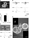
Figure 1.
Figure 1.
VD4 does not innervate supernumerary targets. Identified Lymnaea neurons were isolated in cell culture and paired overnight as soma-soma triplets. Intracellular recordings were made to demonstrate chemical synapses between the paired cells. A, Photomicrograph of a VD4 that was soma-soma paired with two LPeD1 cells. B, Representative traces from pair-wise intracellular recordings of soma-soma triplets. Induced action potentials in VD4 generated 1:1 EPSPs in LPeD1a but not in LPeD1b. C, Summary data showing the percentage of LPeD1a and LPeD1b that formed excitatory synapses with VD4 in CM. D, Phase-contrast picture of soma-soma triplet that was incubated in FM1-43 dye. VD4 was stimulated intracellularly for 2-3 min by injecting pulses of depolarizing current. After dye washout, fluorescent images of FM1-43 loaded cells were acquired. Insets show pseudo-color images of FM1-43, which was localized exclusively in VD4 at its contact site with only one (LPeD1a, top inset) but not the other LPeD1 cell (LPeD1b, bottom inset). E, A photomicrograph depicting cells paired in quadruplet configuration. Specifically, a second VD4 (VD4b) was juxtaposed against a previously uninnervated LPeD1b from a soma-soma triplet. Representative intracellular recordings made 4 hr after juxtaposing the VD4b demonstrate that action potentials in the VD4b cell generate 1:1 EPSPs in LPeD1b. Scale bars: phase-contrast images, 50 μm; fluorescent images, 25 μm.
Figure 2.
Figure 2.
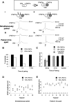
Innervation patterns and synaptic efficacy between VD4 and LPeD1 cells is temporally regulated. To define the precise pattern of synaptic innervation between VD4 and multiple LPeD1 cells, these neurons were juxtaposed against VD4 at different time points. Top (box), experimental paradigm. VD4 was paired with LPeD1a, and the second LPeD1 (LPeD1b) was brought into contact either simultaneously or 1 or 4 hr after contact with the first cell. Pairwise intracellular recordings were made 4 hr after LPeD1b was juxtaposed. When juxtaposed simultaneously, both LPeD1 cells (a and b) were innervated by VD4. Induced action potentials in VD4 generated 1:1 EPSPs in both cells (Ai, Aii). However, when LPeD1b was juxtaposed with VD4 4 hr after LPeD1a, a synapse was detected only in LPeD1a (Aiii, Aiv). B, C, Summarized data of the percentage of synapses formed (B) and absolute EPSP amplitude with each target (C). D, E, Average EPSP amplitude recorded in each postsynaptic target in triplet configuration paired simultaneously (D) or after a 4 hr delay (E). D, EPSP data are shown only for those triplets in which a synapse was formed with both targets.
Figure 3.
Figure 3.
The exclusion of supernumerary target innervation by VD4 is contingent on CM-derived trophic factors. To test whether trophic factors are required for the mediation of exclusion signal, cells were paired in DM. Simultaneous intracellular recordings were made 12-18 hr after pairing. In contrast to cells paired in CM (Fig. 2), in 94% of experiments in which triplets were paired in DM, both LPeD1 cells were innervated simultaneously by VD4; however, these synapses were always inhibitory (inappropriate inhibitory synapses not observed in vivo). A, B, Representative traces from pairwise recording, which show that induced action potentials in VD4 elicit 1:1 IPSPs in both LPeD1 cells. C, Summarized data showing the percentage of inhibitory synapses formed by VD4 with two LPeD1 targets in DM. D, Summary data for average IPSP amplitude measured in each postsynaptic target in soma-soma triplets and control pairs. No significant difference was observed between VD4-induced IPSPs in both LPeD1 cells that were paired in triplet configuration. Similarly, VD4-induced IPSPs in LPeD1 pairs in DM were not significantly different from IPSPs observed in either LPeD1 cell in triplets. E, Summary data for the combined IPSP amplitude of both postsynaptic targets in DM-treated triplets compared with the IPSP amplitude of control pairs. All IPSPs were measured at a postsynaptic membrane potential of -60 mV. Arrows indicate the injection of depolarizing current.
Figure 4.
Figure 4.
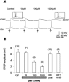
Presynaptic activation of cAMP pathway decreases synaptic efficacy at VD4 and LPeD1 synapses. To test whether cAMP-dependent pathway determines the efficacy of synaptic strength, VD4 was paired with LPeD1 in either absence or presence of 8Br-cAMP. Before intracellular recordings, the drug was washed with normal DM, and the efficacy of synaptic strength was tested. Compared with their control counterparts, increasing concentrations of 8Br-cAMP (10-1000 μm) reduced synaptic efficacy between VD4 and LPeD1. A, Representative traces of intracellular recordings from VD4 and LPeD1. In normal CM, VD4 induced 1:1 EPSPs in LPeD1. However, when cells were maintained overnight in CM containing 8Br-cAMP and subsequently recorded under normal DM conditions, the amplitude of VD4-induced EPSPs decreased in a dose-dependent manner with increasing concentrations of the cAMP analog. To rule out the possibility that postsynaptic responsiveness to ACh (VD4 transmitter) did not change after chronic treatment with 8Br-cAMP, synaptic ACh sensitivity in LPeD1 was tested by exogenously applying ACh to LPeD1 specifically at the contact site with VD4. The response to exogenous ACh was similar in both control and 1000 μm 8Br-cAMP (insets). B, Summary data showing the average EPSP amplitude for all experiments in each condition. Selective treatment of the postsynaptic cell with 8Br-cAMP did not affect EPSP amplitude. Moreover, the reduction of synaptic efficacy in the presence of cAMP analogs was blocked when the presynaptic neurons were coincubated with 8Br-cAMP and H-89.
Figure 5.
Figure 5.
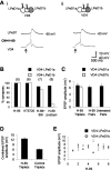
Presynaptic suppression of PKA activity enables hyperinnervation and an enhancement of synaptic efficacy. To test for the involvement of PKA in regulating the synaptic efficacy, cells were paired in the presence of PKA antagonists. Specifically, VD4 was first paired with LPeD1a in the presence of CM + H-89 (PKA antagonist), and, after 4 hr, a second LPeD1 was juxtaposed against VD4. After the drug washout, simultaneous intracellular recordings were made from all cells. A, Representative traces of pairwise recordings made 4 hr after the second LPeD1 was juxtaposed demonstrate that action potentials in VD4 induced 1:1 EPSPs of equal amplitude in both LPeD1 cells.B, Summary data showing the percentage of LPeD1a and LPeD1b neurons that formed synapses with VD4 when the presynaptic neuron was treated selectively with H-89 or KT5720. Hyperinnervation induced by PKA antagonists was not rescued by the addition of the cAMP analog 8Br-cAMP. In control experiments, VD4 innervated only one postsynaptic target when the postsynaptic neuron was treated selectively with H-89. C, After H-89 pretreatment, the average EPSP amplitude for each LPeD1 neuron, either in a triplet configuration or when paired, is compared with the control cells. No significant difference was observed between VD4-induced EPSPs in both LPeD1 cells that were paired with a single VD4 in the presence of H-89. Similarly, VD4-induced EPSPs in LPeD1 pairs in H-89 was identical to that of control pairs and also to those cultured as triplets in the presence of PKA inhibitor. D, Summary data for the combined EPSP amplitude of both postsynaptic targets in H-89-treated triplets compared with the total EPSP amplitude of control triplets, in which only one postsynaptic target was innervated. E, Scatterplot of the average EPSP amplitude for both postsynaptic targets in H-89-treated triplets.
Figure 6.
Figure 6.
cAMP analogs rescue hyperinnervation in the absence of trophic factors. VD4-LPeD1 triplets were paired in the absence of trophic factors and treated with 8Br-cAMP (100 μm). A, Pairwise intracellular recordings revealed that VD4 innervated only one LPeD1 target. Ai, Evoked action potentials in VD4 induced 1:1 IPSPs in LPeD1a; in Aii, no postsynaptic response was detected in LPeD1b. B, Summary data of the percentage of LPeD1a and LPeD1b neurons that were innervated by VD4 neurons pretreated with PKA agonists. All postsynaptic responses were measured at a postsynaptic membrane potential of -60 mV. Arrows indicate the injection of depolarizing current.

Similar articles

Cited by

References

    1. Ahmari SE, Smith SJ ( 2002) Knowing a nascent synapse when you see it. Neuron 34: 333–336. - PubMed
    1. Casadio A, Martin KC, Giustetto M, Zhu H, Chen M, Bartsch D, Bailey CH, Kandel ER ( 1999) A transient, neuron-wide form of CREB-mediated long-term facilitation can be stabilized at specific synapses by local protein synthesis. Cell 99: 221–237. - PubMed
    1. Colledge M, Dean RA, Scott GK, Langeberg LK, Huganir RL, Scott JD ( 2000) Targeting of PKA to glutamate receptors through a MAGUK-AKAP complex. Neuron 27: 107–119. - PubMed
    1. Colman H, Nabekura J, Lichtman JW ( 1997) Alterations in synaptic strength preceding axon withdrawal. Science 275: 356–361. - PubMed
    1. Davis GW, Bezprozvanny I ( 2001) Maintaining the stability of neural function: a homeostatic hypothesis. Annu Rev Physiol 63: 847–869. - PubMed

Publication types

MeSH terms