. 2003 May 15;23(10):4219-27.
doi: 10.1523/JNEUROSCI.23-10-04219.2003.

Delayed systemic Nogo-66 receptor antagonist promotes recovery from spinal cord injury

Affiliations

Delayed systemic Nogo-66 receptor antagonist promotes recovery from spinal cord injury

Shuxin Li et al. J Neurosci. .

Abstract

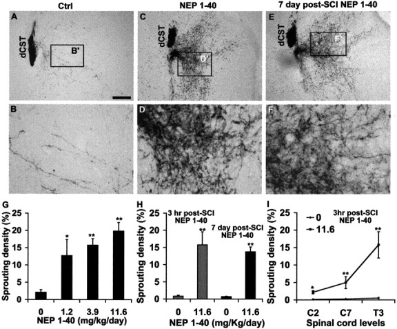
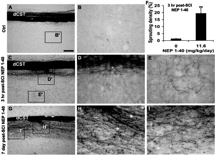
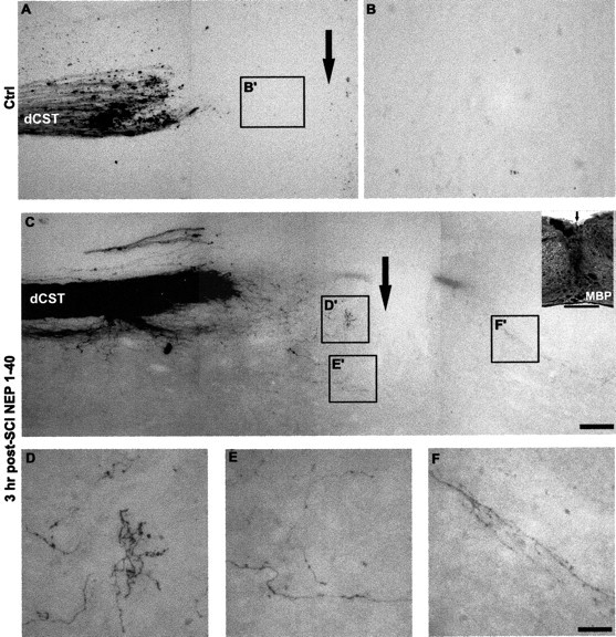
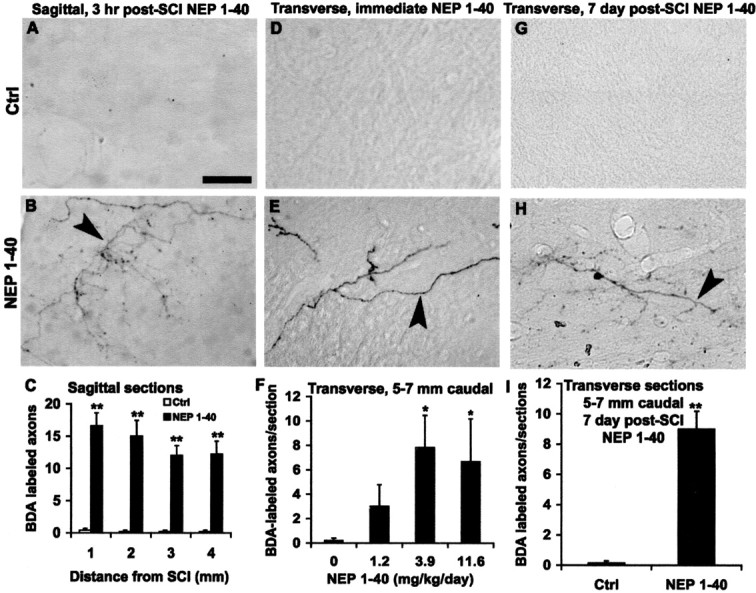
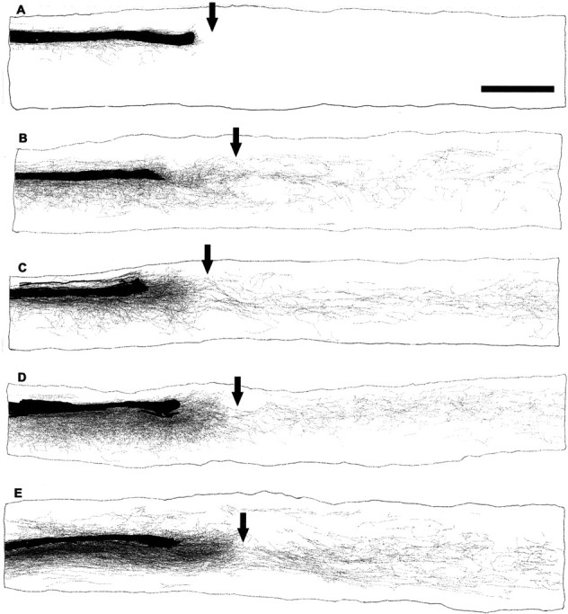
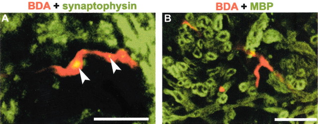
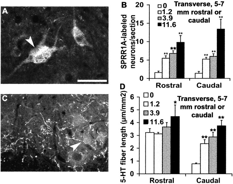
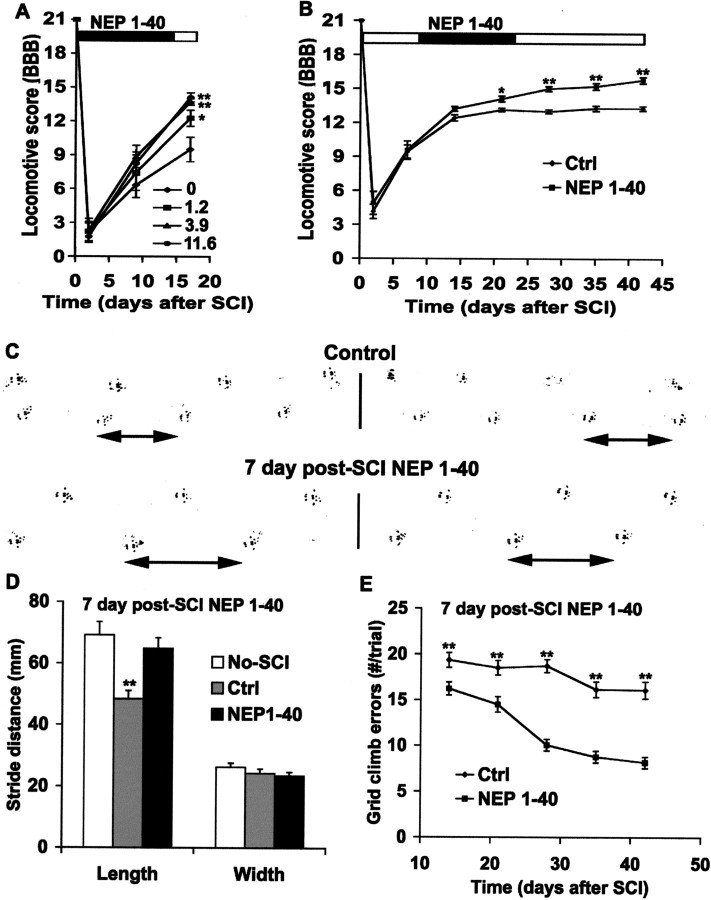
Traumatized axons possess an extremely limited ability to regenerate within the adult mammalian CNS. The myelin-derived axon outgrowth inhibitors Nogo, oligodendrocyte-myelin glycoprotein, and myelin-associated glycoprotein, all bind to an axonal Nogo-66 receptor (NgR) and at least partially account for this lack of CNS repair. Although the intrathecal application of an NgR competitive antagonist at the time of spinal cord hemisection induces significant regeneration of corticospinal axons, such immediate local therapy may not be as clinically feasible for cases of spinal cord injury. Here, we consider whether this approach can be adapted to systemic therapy in a postinjury therapeutic time window. Subcutaneous treatment with the NgR antagonist peptide NEP1-40 (Nogo extracellular peptide, residues 1-40) results in extensive growth of corticospinal axons, sprouting of serotonergic fibers, upregulation of axonal growth protein SPRR1A (small proline-rich repeat protein 1A), and synapse re-formation. Locomotor recovery after thoracic spinal cord injury is enhanced. Furthermore, delaying the initiation of systemic NEP1-40 administration for up to 1 week after cord lesions does not limit the degree of axon sprouting and functional recovery. This indicates that the regenerative capacity of transected corticospinal tract axons persists for weeks after injury. Systemic Nogo-66 receptor antagonists have therapeutic potential for subacute CNS axonal injuries such as spinal cord trauma.


Figures

Figure 1.
Figure 1.
Systemic NEP1–40 promotes dCST sprouting in transverse sections rostral to the dorsal hemisection of the spinal cord. A, B, The BDA-labeled dorsal CST is visualized in a transverse section 5–7 mm rostral to the hemisection as a discrete bundle in a vehicle-treated animal. Several BDA-labeled fibers project into the gray matter area. Dorsal is up in this and all transverse sections.C,D,A transverse section 5–7 mm rostral to the hemisection from a NEP1–40-treated animal(subcutaneous administration via minipump) exhibits a high density of sprouts extending laterally into the gray matter, some of them into the white matter area.E,F,A transverse section 5–7 mm rostral to the hemisection from an animal receiving NEP1–40 treatment 7 d after hemisection (subcutaneous administration via minipump) exhibits an identical dCST sprouting pattern as for immediate peptide treatment. B′, D′, and F′ are shown at higher magnification in B, D, and F, respectively. G, The sprouting density of BDA-labeled fibers outside of the dorsal CST is illustrated for several doses of immediate subcutaneous NEP1–40 treatment mice. H, Bar graph indicates the sprouting density of BDA-labeled fibers outside of the dorsal CST from 3 hr intraperitoneal delayed treatment in mice (gray bars) and 7 d subcutaneous delayed administration in mice (black bars). I, The number of ectopic CST sprouts at the C2, C7, and T3 spinal levels from 3 hr intraperitoneal posttreatment animals. Means ± SEM from seven mice in each group are reported. Scale bar:(inA)A,C,E,100μm;B,D,F,25μm. The NEP1–40 values are statistically different from control. *p< 0.05; **p < 0.01; Student's t test.
Figure 2.
Figure 2.
Delayed systemic NEP1–40 stimulates CST sprouting in rostral parasagittal sections. Sections 1–4 mm rostral to the dorsal hemisection of the spinal cord from the control group (A, B) reveal the longitudinal dCST (A) and few BDA-labeled fibers in the gray matter (B). Many collateral sprouts with a branching pattern are seen in the sections receiving intraperitoneal NEP1–40 treatment 3 hr after injury (C–E). Quantification of sprouting from longitudinal sections 1–4 mm rostral to spinal cord lesion indicates that the sprouting density of dorsal CST in the animals receiving NEP1–40 3 hr after treatment is more than 10 times greater than that in the time-matched controls (F). Longitudinal sections (G–I) from mice receiving NEP1–40 treatment 7 d after hemisection illustrate a similar spouting pattern as for 3 hr delayed drug delivery. In all the sagittal sections rostral is to the left and dorsalisup.B′,D′,E′,I′, and H′ are shown at higher magnification in B,D,E,I, and H, respectively. Scale bar: (inA) A,C,G,100μm; B, D, E, H, I,25μm. Means ± SEM from seven mice in each group are reported. The NEP1–40 values are statistically different from control. **p < 0.01; Student's t test.
Figure 3.
Figure 3.
Systemic NEP1–40 promotes dCST growth through the SCI site. A,B,A parasagittal section containing the transection site (arrow) from a vehicle-treated animal demonstrates the transection of BDA-labeled dCST fibers at the injury site. No fibers extend into the area of the trauma or beyond it. C, A section from an animal receiving intraperitoneal NEP1–40 demonstrates a similar degree of dorsal hemisection injury, but numerous branched, sprouting fibers are observed in the areas around the transection site. The higher magnifications of these areas in D–F′ illustrate the meandering course of the regenerating CST fibers in the NEP1–40-treated animals (D–F). The inset in C immunostained for MBP illustrates the disruption of staining in the hemisection site. Scale bars: (in C) A, C, 100 μm (A, C); (in F) B, D–F, 25 μm; inset, 500 μm.
Figure 5.
Figure 5.
Systemic NgR antagonist peptide induces numerous distal regenerating CST fibers. A, B, Parasagittal sections 1–4 mm distal to the hemisection display the numerous branching fibers (arrowhead) in the peptide-treated mice (B), in contrast to no fibers in the control sections (A). C, Quantification of CST fibers is illustrated at various distances caudal to the injury site for the group given treatment 3 hr after trauma. D, E, Transverse sections at a level 5–7 mm caudal to the lesion illustrate no fibers in the gray matter of the dorsal spinal cord from the vehicle-treated animals (D), but a large number of fibers with branching patterns in the NEP1–40 group are seen (arrowhead) ventral to dorsal CST (E). F, CST fiber counts at a level of 5–7 mm caudal to the lesion from transverse sections indicate a greater number in NEP1–40-treated groups (subcutaneous application) than in the control group. G, H, Transverse sections 5–7 mm caudal to the lesion show a number of BDA-labeled fibers (H, arrowhead) from mice treated with NEP1–40 7 d after SCI delivery, but no fibers in the sections from the time-matched vehicle-treated animals (G). I, Graph shows the number of BDA-labeled CST fibers 5–7 mm distal to hemisection from animals receiving NEP1-40 treatment 7 d after injury. Scale bar, 25 μm. In all the graphs, means ± SEM from seven mice in each group are reported. The NEP1–40 values are statistically different from the control. *p < 0.05; **p < 0.01; Student's t test.
Figure 4.
Figure 4.
Camera lucida reconstructions of consecutive parasagittal sections around the lesion site. The injury site is indicated by an arrow. Control animals show no regenerative growth (A). In contrast, NEP1–40 treatment 3 hr (B–D) or 7 d (E) after trauma induces a high density of sprouting of the lesioned dorsal CST fibers. Some of these regenerated fibers grow around and through the lesion, and project into the distal spinal cord, particularly into the gray matter areas. Scale bar, 1 mm.
Figure 6.
Figure 6.
Synaptophysin and MBP distribution relative to caudal CST fibers in NEP1–40-treated mice. A, Confocal imaging displays the double-staining for BDA-labeled regenerating CST fibers (red) and synaptophysin (green) in the ventral horn of transverse spinal cord at a level of 8–9 mm caudal to the lesion site from NEP1–40-treated mice. Note the localization of synaptophysin to varicosities (arrowheads) of BDA-positive fibers in the lumbar level of spinal cord.B, Transverse section of ventral gray matter 8–9 mm caudal to the hemisection stained for BDA (red) and MBP (green) illustrates that NEP1–40-induced CST fibers in the distal spinal cord are not myelinated 20 d after SCI. Scale bars, 25 μm.
Figure 7.
Figure 7.
Systemic administration of NEP1–40 induces an upregulation of SPRR1A and distal serotonin fiber sprouting. A, A ventral horn transverse section from 5–7 mm caudal to the lesion site displays the positive neurons (arrowhead) stained with regeneration-related protein SPRR1A from NEP1–40-treated animals. B, SPRR1A-positive neuronal counts at a level of 5–7 mm rostral or 5–7 mm caudal to the lesion demonstrate a significant increase in the number of immunoreactive cells in the NEP1–40-treated groups. C, A transverse section of ventral horn 5–7 mm distal to the hemisection illustrates the serotonin fibers stained with antibody against 5-HT (arrowhead). D, Immunoreactive serotonin fiber length in the ventralhorn at a level of 5–7 mm rostral or 5–7 mm caudal to injury site was measured. The means ± SEM from seven mice in each group are reported. The NEP1–40 values are statistically different from the control. *p < 0.05; **p < 0.01; Student's t test. Scale bar: (in A) A, C, 25 μm.
Figure 8.
Figure 8.
Systemic NgR antagonist peptide improves functional recovery after dorsal hemisection. A, The locomotor BBB score is reported as a function of time after dorsal hemisection in the vehicle-or NEP1–40-treated animals initiated at the time of SCI. B, The BBB score is plotted as a function of time after SCI in control or NEP1–40 animals receiving treatment 7 d after trauma. C, Two examples of the representative footprints from control (top) or NEP1–40 (bottom) animals treated 7 d after SCI are shown. The double-headed arrows indicate strike length for one step. D, Footprint analysis reveals a shorter stride length in control mice than uninjured or injured + NEP1–40 mice(treatment 7 d after injury), where as no differences are found in the stride width among the three groups. E, Hindlimb errors during inclined grid climbing are reported as a function of post-SCI time in the control or NEP1–40-treated groups (treatment after 7 d). In all the graphs, means± SEM from seven mice in each group are reported. The NEP1–40 values are statistically different from the control in A, B, and E. The control values are statistically different from no-SCI or SCI + NEP1–40 mice in D. *p < 0.05; **p < 0.01; Student's t test.

Similar articles

Cited by

References

    1. Bartsch U, Bandtlow CE, Schnell L, Bartsch S, Spillmann AA, Rubin BP, Hillenbrand R, Montag D, Schwab ME, Schachner M ( 1995) Lack of evidence that myelin-associated glycoprotein is a major inhibitor of axonal regeneration in the CNS. Neuron 15: 1375–1381. - PubMed
    1. Basso DM, Beattie MS, Bresnahan JC, Anderson DK, Faden AI, Gruner JA, Holford TR, Hsu CY, Noble LJ, Nockels R, Perot PL, Salzman SK, Young W ( 1996) MASCIS evaluation of open field locomotor scores: effects of experience and teamwork on reliability. Multicenter Animal Spinal Cord Injury Study. J Neurotrauma 13: 343–359. - PubMed
    1. Bomze HM, Bulsara KR, Iskandar BJ, Caroni P, Skene JH ( 2001) Spinal axon regeneration evoked by replacing two growth cone proteins in adult neurons. Nat Neurosci 4: 38–43. - PubMed
    1. Bonilla IE, Tanabe K, Strittmatter SM ( 2002) Small proline-rich repeat protein 1A is expressed by axotomized neurons and promotes axonal outgrowth. J Neurosci 22: 1303–1315. - PMC - PubMed
    1. Bradbury EJ, Moon LD, Popat RJ, King VR, Bennett GS, Patel PN, Fawcett JW, McMahon SB ( 2002) Chondroitinase ABC promotes functional recovery after spinal cord injury. Nature 416: 636–640. - PubMed

Publication types

MeSH terms