. 2003 May 15;23(10):4378-85.
doi: 10.1523/JNEUROSCI.23-10-04378.2003.

Variable dopamine release probability and short-term plasticity between functional domains of the primate striatum

Affiliations

Variable dopamine release probability and short-term plasticity between functional domains of the primate striatum

Stephanie J Cragg. J Neurosci. .

Abstract

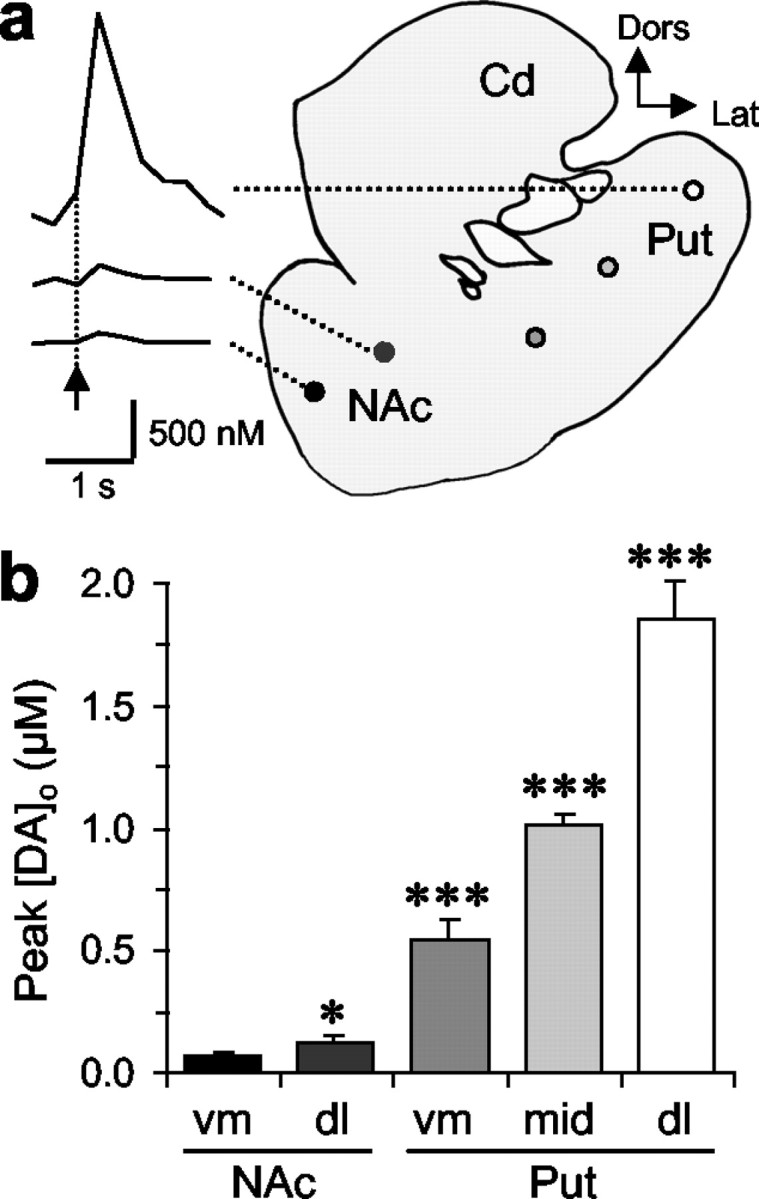
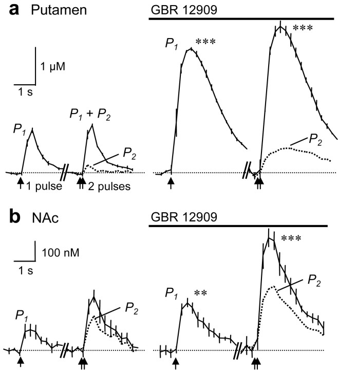
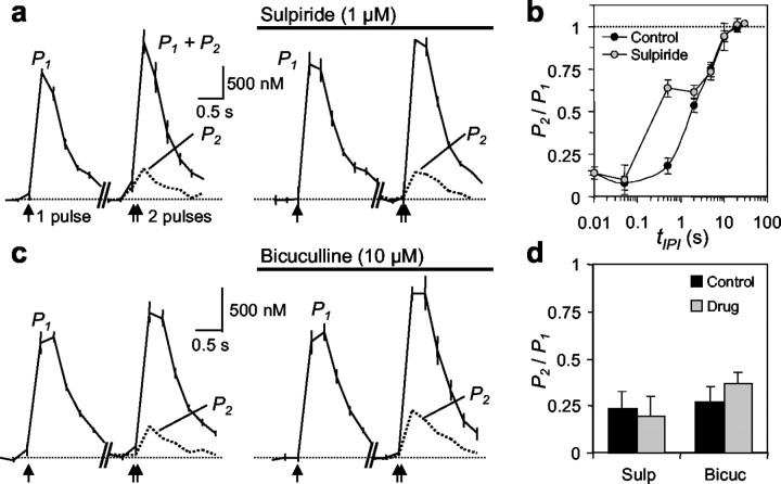
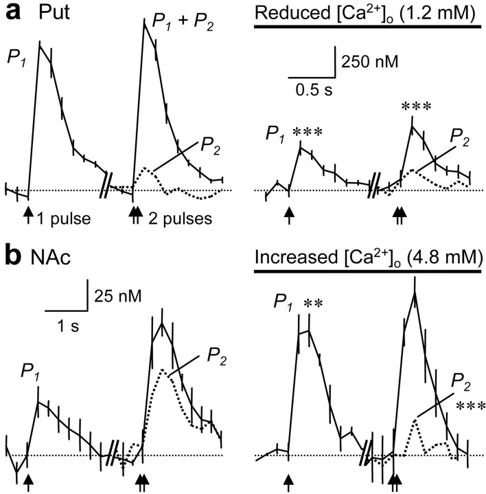
Release of the neuromodulator dopamine (DA) is critical to the control of locomotion, motivation, and reward. However, the probability of DA release is not well understood. Current understanding of neurotransmitter release probability in the CNS is limited to the conventional synaptic amino acid transmitters (e.g., glutamate and GABA). These fast neurotransmitters are released with a repertoire of probabilities according to synapse type, and these probabilities show activity-dependent plasticity according to synapse use. Synapses for neuromodulators such as DA, however, are designed for signaling that diverges temporally and spatially from that for fast neurotransmitters: DA receptors are exclusively metabotropic and at sites that extend to extrasynaptic locations and neighboring synapses. In this study, the release probability of DA was explored in real time in limbicversus motor-associated functional domains of the striatum of a primate (marmoset; Callithrix jacchus) using fast-scan voltammetry at a carbon-fiber microelectrode. We show that the probability of axonal DA release varies with striatal domain. Furthermore, release probability exhibits a short-term, activity-dependent plasticity that ranges from depression to facilitation in motor-through limbic-associated regions, respectively. Rapid plasticity does not result from metabotropic D2-like DA receptor activation or ionotropic GABA(A) receptor effects but is dependent on Ca2+ availability. These data reveal that rapid dynamics in DA release probability will participate in the transmission of the patterns and frequencies encoded by DA neuron action potential discharge. Furthermore, the regional variation in these features indicates that limbic-versus motor-associated DA neurons are permitted to generate diverse DA signals in response to a given firing pattern.


Figures

Figure 1.
Figure 1.
Cyclic voltammograms and uptake transport identify DA. a, Cyclic voltammograms obtained in putamen (Put; dorsolateral; gray) and, at 6× amplification, NAc (lateral; black). Scan range, −0.7 to +1.3 to −0.7 V. Dashed lines indicate oxidation (asterisk) and reduction peaks for DA. Calibrations: putamen, 6 nA; NAc, 1 nA. b, c, Mean [DA]o ± SEM versus time caused by a single pulse (arrow) in NAc (ventromedial) in control (circles) and during inhibition of either the NET [desipramine (Desip; 300 nm); triangles] (b) or DAT [GBR 12909 (GBR; 500 nm); squares] (c). b, Desipramine had no effect on removal rate or [DA]o (n = 6). c, GBR 12909 inhibited removal and enhanced [DA]o (***p < 0.001; n = 5).
Figure 2.
Figure 2.
Availability of DA for release from ventral through dorsal striatum. a, Typical observations of [DA]o versus time after one stimulus pulse (arrow) in NAc versus dorsolateral putamen (Put) in coronal sections of marmoset striatum. Cd, Caudate; Dors, dorsal; Lat; lateral. Calibration for [DA]o: 500 nm, 1 sec. Filled circles are site-specific and indicate sites of recording for mean data, corresponding to the filled bar in b.b, Mean peak [DA]o ± SEM evoked at five loci indicated in a, along a ventromedial–dorsolateral axis. DA release varies between and within each region of the ventral and dorsal striatum (two-way ANOVA; p < 0.001). Post hoc comparisons with ventromedial NAc are illustrated (*p < 0.05; ***p < 0.001; n = 7–27).
Figure 3.
Figure 3.
Short-term plasticity and striatal domain. a, b, Mean [DA]o ± SEM (solid lines with hatching) versus time evoked (arrows) by a single pulse (P1) (left) or two pulses (P1 + P2) paired at 10 msec (100 Hz), administered when P1 is reproducible (right) (see Materials and Methods). The release caused by the second pulse (P2) (dotted plot) is determined in all measurements by subtraction of P1 from P1 + P2. a, In putamen (dl), a marked PPD is prominent (P2 vs P1; p < 0.001; n = 11–15). Calibration: 250 nm DA, 0.5 sec. b, In contrast, in NAc (vm), there is PPF (P2 vs P1; p < 0.001; n = 20–23). Calibration: 50 nm DA, 0.5 sec. c, The paired-pulse ratio (P2/P1) ± SEM at loci throughout the ventromedial–dorsolateralstriatal extentis inversely related to P1 (y = ax−b; a = 0.16; b = 1.07; R2 > 0.99; solid line; n = 7–23). A similar relationship remains when P2 is normalized for the regional, threefold variation in tissue DA content (Cragg et al., 2000) (dashed line; R2 = 0.97; a = 0.03; b = 1.8). Dorsal striatum is associated with PPD, and ventral striatum is associated with PPF (parity; dotted line). Filled circles correspond to recording sites as indicated in Figure 2a. d, Paired-pulse plasticity ± SEM varies dynamically with tIPI (n = 5–20). PPF in NAc decays at a tIPI of >50 msec. PPD is immediate in dorsal striatum and residual in NAc (lateral) after tIPI = 50 msec; the paired-pulse ratio recovers to parity (dotted line) over time by an exponential relationship in NAc (data not shown) as in putamen (Put) (Cragg et al., 2002). Ventromedial NAc is not plotted between tIPI of 50 msec and <5 sec, because a PPD (projected; dashed line) prevents accurate quantification of the low [DA]o in this region until >5 sec.
Figure 4.
Figure 4.
Variable short-term plasticity is not attributable to uptake. a, b, Mean [DA]o ± SEM (solid lines with hatching) versus time evoked (arrows) by P1, P1 + P2 (tIPI = 10 msec), and P2 (dotted plot) in putamen (dorsolateral) (a) and NAc (ventromedial) (b) during control (left) and DAT inhibition [GBR 12909 (500 nm); solid bar] (right). a, In putamen, DAT inhibition enhances lifetime and peak [DA]o of P1 and P1 + P2 compared with control (***p<0.001), but the PPD is not modified (n = 5–6). Calibration: 1μm DA, 1 sec. b, In NAc, DAT inhibition enhances lifetime and peak [DA]o of P1 and P1 + P2 compared with control (**p < 0.01; ***p < 0.001), but the PPF is not significantly modified (n = 5–6). Calibration: 100 μm DA, 1 sec.
Figure 5.
Figure 5.
Eliminating variable input activity as a component of PPD. a, c, Mean [DA]o ± SEM (solid lines with hatching) versus time in putamen (dorsolateral) evoked (arrows) by a single pulse (P1), paired pulses (P1 + P2), and a second pulse (P2; dotted plot) (tIPI = 10 msec), during control (left) and antagonism of D2-like DA receptors [sulpiride (1 μm)] (a) or GABAA receptors [bicuculline (10 μm)] (c) (solid bars) (right). Calibration: 500 nm DA, 0.5 sec. a, Sulpiride did not modify P1, P1 + P2, or P2 compared with control at this tIPI (n = 3–6). b, Paired-pulse ratios ± SEM versus tIPI in central (filled circles) and with sulpiride (graycircles) tIPI is plotted logarithmically. The dynamic variation in PPD with tIPI is modified by D2 receptors only at >50msec and <2 sec (*p < 0.001). Dotted line indicates P2 = P1. c, Bicuculline did not modify P1, P1 + P2, or P2 compared with control (n = 6–7). d, Paired-pulse ratios (P2 /P1) ± SEM for each control (filled) versus drug treatment (shaded) in a and c. Sulp, Sulpiride; Bicuc, bicuculline.
Figure 6.
Figure 6.
Modifying initial release probability alters short-term plasticity. Mean [DA]o ± SEM (solid lines with hatching) versus time evoked (arrows) by a single pulse (P1), paired pulses (P1 + P2) and a second pulse (P2; dotted plot) (tIPI = 10 msec) in putamen (Put; dorsolateral) (a) and NAc (ventromedial) (b) during control (left) and manipulation of [Ca2+]o (solid bar) (right). a, In putamen, halving [Ca2+]o to 1.2 mM reduces P1 and P1 + P2 compared with controls (***p < 0.001; n = 4) but significantly enhances P2 /P1 (**p < 0.01). A significant PPD remains (p < 0.01; P2 vs P1). Calibration: 250 nm DA, 0.5 sec. b, In NAc, doubling [Ca2+]o to 4.8 mm enhances P1 compared with control (**p < 0.01; n = 7) but not P1 + P2. P2 is markedly reduced (***p < 0.001), and PPF is significantly reduced (***p < 0.001) to a significant PPD (p < 0.001; P2 vs P1). Calibration: 25 nm DA, 1 sec.

References

    1. Abeliovich A, Schmitz Y, Farinas I, Choi-Lundberg D, Ho WH, Castillo PE, Shinsky N, Verdugo JM, Armanini M, Ryan A, Hynes M, Phillips H, Sulzer D, Rosenthal A ( 2000) Mice lacking alpha-synuclein display functional deficits in the nigrostriatal dopamine system. Neuron 25: 239–252. - PubMed
    1. Atwood HL, Karunanithi S ( 2002) Diversification of synaptic strength: presynaptic elements. Nat Rev Neurosci 3: 497–516. - PubMed
    1. Auger C, Kondo S, Marty A ( 1998) Multivesicular release at single functional synaptic sites in cerebellar stellate and basket cells. J Neurosci 18: 4532–4547. - PMC - PubMed
    1. Avshalumov MV, Chen BT, Marshall SP, Pena DM, Rice ME ( 2003) Glutamate-dependent inhibition of dopamine release in striatum is mediated by a new diffusible messenger, H2O2 J Neurosci 23: 2744–2750. - PMC - PubMed
    1. Bellingham MC, Walmsley B ( 1999) A novel presynaptic inhibitory mechanism underlies paired pulse depression at a fast central synapse. Neuron 23: 159–170. - PubMed

Publication types

MeSH terms

LinkOut - more resources