

Molecular characterization of the skate peripherin/rds gene: relationship to its orthologues and paralogues
- PMID: 12766040
- PMCID: PMC2991160
- DOI: 10.1167/iovs.02-1152
Molecular characterization of the skate peripherin/rds gene: relationship to its orthologues and paralogues
Abstract
Purpose: A great deal of information about functionally significant domains of a protein may be obtained by comparison of primary sequences of gene homologues over a broad phylogenetic base. This study was designed to identify evolutionarily conserved domains of the photoreceptor disc membrane protein peripherin/rds by analysis of the homologue in a primitive vertebrate, the skate.
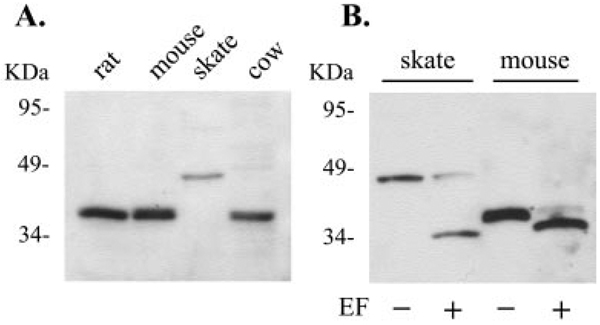
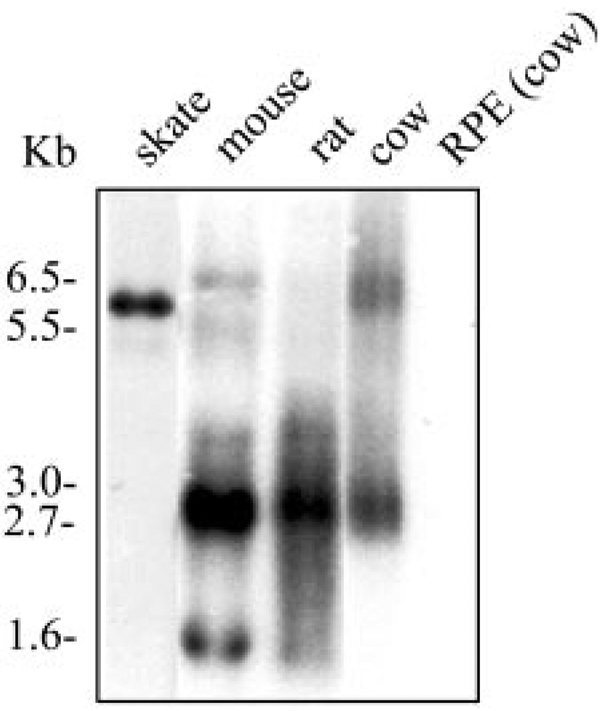
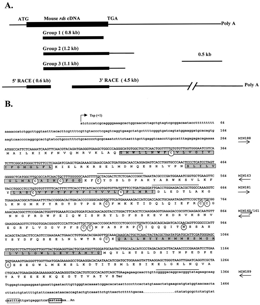
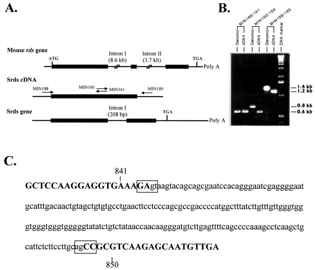
Methods: A skate retinal cDNA library was screened using a mouse peripherin/rds clone. The 5' and 3' untranslated regions of the skate peripherin/rds (srds) cDNA were isolated by the rapid amplification of cDNA ends (RACE) approach. The gene structure was characterized by PCR amplification and sequencing of genomic fragments. Northern and Western blot analyses were used to identify srds transcript and protein, respectively.
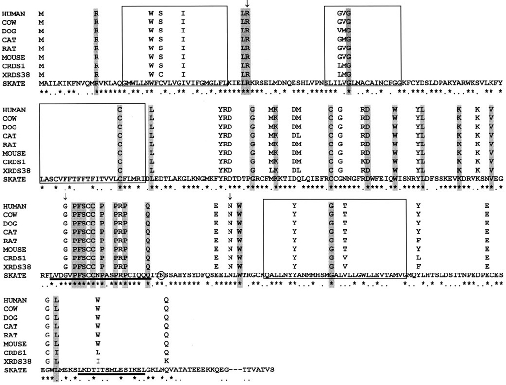
Results: A new homologue of peripherin/rds was identified from the skate retinal cDNA library. SRDS is a glycoprotein with a predicted molecular mass of 40.2 kDa. The srds gene consists of two exons and one small intron and transcribes into a single 6-kb message. Phylogenetic analysis places SRDS at the base of peripherin/rds family and near the division of that group and the branch leading to rds-like and rom-1 genes. SRDS protein is 54.5% identical with peripherin/rds across species. Identity is significantly higher (73%) in the intradiscal domains. Sequence comparison revealed the conservation of all residues that have been shown, on mutation, to associate with retinitis pigmentosa and showed conservation of most residues associated with macular dystrophies. Comparison with ROM-1 and other rds-like proteins revealed the presence of a highly conserved domain in the large intradiscal loop.
Conclusions: Srds represents the skate orthologue of mammalian peripherin/rds genes. Conservation of most of the residues associated with human retinal diseases indicates that these residues serve important functional roles. The high degree of conservation of a short stretch within the large intradiscal loop also suggests an important function for this domain.
Figures

References
-
- Steinberg RH, Fisher SK, Anderson DH. Disc morphogenesis in vertebrate photoreceptors. J Comp Neurol. 1980;190:501–518. - PubMed
-
- Boesze-Battaglia K, Lamba OP, Napoli AA, Jr, Sinha S, Guo Y. Fusion between retinal rod outer segment membranes and model membranes: a role for photoreceptor peripherin/rds. Biochemistry. 1998;37:9477–9487. - PubMed
-
- Ma J, Norton JC, Allen AC, et al. Retinal degeneration slow (rds) in mouse results from simple insertion of a t haplotype-specific element into protein-coding exon II. Genomics. 1995;28:212–219. - PubMed
-
- Cheng T, Al-Ubaidi MR, Naash MI. Structural and developmental analysis of the mouse peripherin/rds gene. Somat Cell Mol Genet. 1997;23:165–183. - PubMed
Publication types
MeSH terms
Substances
Grants and funding
LinkOut - more resources
Full Text Sources
