Case Reports
. 2003 Jun 10;100(12):7377-82.
doi: 10.1073/pnas.1230273100. Epub 2003 May 23.

Myasthenic syndrome caused by mutation of the SCN4A sodium channel

Affiliations
Case Reports

Myasthenic syndrome caused by mutation of the SCN4A sodium channel

Akira Tsujino et al. Proc Natl Acad Sci U S A. .

Abstract

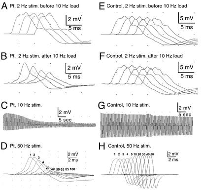
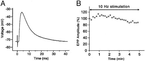
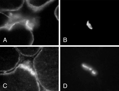
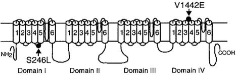
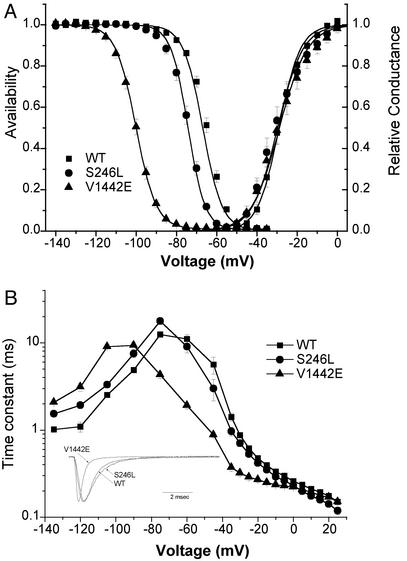
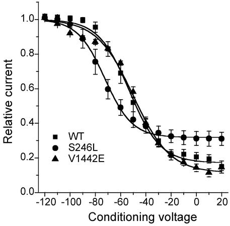
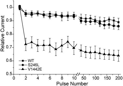
In a myasthenic syndrome associated with fatigable generalized weakness and recurrent attacks of respiratory and bulbar paralysis since birth, nerve stimulation at physiologic rates rapidly decremented the compound muscle action potential. Intercostal muscle studies revealed no abnormality of the resting membrane potential, evoked quantal release, synaptic potentials, acetylcholine receptor channel kinetics, or endplate ultrastructure, but endplate potentials depolarizing the resting potential to -40 mV failed to excite action potentials. Pursuing this clue, we sequenced SCN4A encoding the skeletal muscle sodium channel (Nav1.4) and detected two heteroallelic mutations involving conserved residues not present in 400 normal alleles: S246L in the S4/S5 cytoplasmic linker in domain I, and V1442E in the S3/S4 extracellular linker in domain IV. The genetically engineered V1442E-Na channel expressed in HEK cells shows marked enhancement of fast inactivation close to the resting potential, and enhanced use-dependent inactivation on high-frequency stimulation; S246L is likely a benign polymorphism. The V1442E mutation in SCN4A defines a novel disease mechanism and a novel phenotype with myasthenic features.


Figures

Fig. 1.
Fig. 1.
Hypothenar CMAP in patient (AD) and thenar CMAP in control (EH) evoked by stimulation at 2, 10, and 50 Hz. Ten-hertz stimulation for 5 min (C) or 50-Hz stimulation for 2 sec (D) decrement the patient CMAP by ≈85%; 2-Hz stimulation for 2 sec has no effect on the patient CMAP unless it is preceded by a 10-Hz conditioning train (A and B). Control CMAP decrements slightly after 10-Hz stimulation (G), pseudofacilitates after 50-Hz stimulation (H), and is unaffected by 2-Hz stimulation before or after a 10-Hz conditioning train (E and F). Numbers in D and H indicate nth CMAPs elicited during 50-Hz stimulation.
Fig. 2.
Fig. 2.
(A) EPP recorded from patient intercostal muscle depolarizes the resting potential from -73 to -43 mV but fails to elicit an action potential. (B) The EPP elicited by 10-Hz nerve stimulation over 5 min first facilitates and then decreases by -13% from baseline. Individual symbols represent means of 100 EPPs recorded over 10-sec intervals.
Fig. 3.
Fig. 3.
Sodium channel (A and C) and AChE (B and D) localization in patient (A and B) and control (C and D) muscle fibers. EPs are identified by localization of AChE in the same sections. Sodium channel expression is similar at the surface membrane and is similarly enhanced at the EP in patient and control fibers. (Magnification, ×370.)
Fig. 4.
Fig. 4.
Scheme of skeletal muscle sodium channel Nav1.4 encoded by SCN4A and the identified mutations.
Fig. 5.
Fig. 5.
Gating behavior of heterologously expressed sodium channels. (A) Channel availability (left) after a 300-msec prepulse was left-shifted for mutant channels, whereas activation (right) was unchanged. Parameter values for Boltzmann fits are listed in Table 3. (B) The kinetics of fast-inactivation were left-shifted for mutant channels (n - 7–12). Inactivation kinetics were measured as: recovery at -135 to -80 mV after 30-msec prepulse to -10 mV, closed-state entry with progressively longer prepulses over a range from -60 to -45 mV, or as the current decay rate for test depolarization ≥ -45 mV. For V1442E, the recovery/prepulse voltages for the inactivation protocols were shifted by -30 mV, to detect Na current in the presence of the large left-shift of inactivation. (Inset) Superposition of amplitude-normalized responses for a depolarization to -30 mV from a holding potential of -120 mV.
Fig. 6.
Fig. 6.
Slow inactivation after a 30-sec conditioning pulse, and an intervening recovery at -120 mV for 20 msec to remove fast inactivation, was less complete for S246L and shifted toward more negative potentials (see Table 3 for fitted parameters).
Fig. 7.
Fig. 7.
Relative Na current during a 50-Hz train of 3-msec depolarizations to -10 mV from a holding potential of -100 mV. Every 20th response is shown after the break at the 10th pulse.

References

    1. Salpeter, M. M. (1987) in Vertebrate Neuromuscular Junctions: General Morphology, Molecular Organization, and Functional Consequences, ed. Salpeter, M. M. (Liss, New York), pp. 1-54.
    1. Flucher, B. E. & Daniels, M. P. (1989) Neuron 3, 163-175. - PubMed
    1. Ruff, R. L. (1996) Acta Physiol. Scand. 156, 159-168. - PubMed
    1. Martin, A. R. (1994) Proc. R. Soc. London B 258, 321-326. - PubMed
    1. Wood, S. J. & Slater, C. P. (2001) Prog. Neurobiol. 64, 393-429. - PubMed

Publication types

Associated data