Cloning of a salivary gland metalloprotease and characterization of gelatinase and fibrin(ogen)lytic activities in the saliva of the Lyme disease tick vector Ixodes scapularis
- PMID: 12767911
- PMCID: PMC2903890
- DOI: 10.1016/s0006-291x(03)00857-x
Cloning of a salivary gland metalloprotease and characterization of gelatinase and fibrin(ogen)lytic activities in the saliva of the Lyme disease tick vector Ixodes scapularis
Abstract
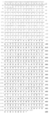
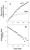
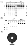
The full-length sequence of tick salivary gland cDNA coding for a protein similar to metalloproteases (MP) of the reprolysin family is reported. The Ixodes scapularis MP is a 488 amino acid (aa) protein containing pre- and pro-enzyme domains, the zinc-binding motif HExxHxxGxxH common to metalloproteases, and a cysteine-rich region. In addition, the predicted amino-terminal sequences of I. scapularis MPs were found by Edman degradation of PVDF-transferred SDS/PAGE-separated tick saliva proteins, indicating that these putative enzymes are secreted. Furthermore, saliva has a metal-dependent proteolytic activity towards gelatin, fibrin(ogen), and fibronectin, but not collagen or laminin. Accordingly, I. scapularis saliva has a rather specific metalloprotease similar to the hemorrhagic proteases of snake venoms. This is the first description of such activity in tick saliva and its role in tick feeding and Borrelia transmission is discussed.
Figures


Similar articles
-
Exploring the sialome of the tick Ixodes scapularis.J Exp Biol. 2002 Sep;205(Pt 18):2843-64. doi: 10.1242/jeb.205.18.2843. J Exp Biol. 2002. PMID: 12177149
-
Purification of a serine protease and evidence for a protein C activator from the saliva of the tick, Ixodes scapularis.Toxicon. 2014 Jan;77:32-9. doi: 10.1016/j.toxicon.2013.10.025. Epub 2013 Oct 31. Toxicon. 2014. PMID: 24184517 Free PMC article.
-
Reprolysin metalloproteases from Ixodes persulcatus, Rhipicephalus sanguineus and Rhipicephalus microplus ticks.Exp Appl Acarol. 2014 Aug;63(4):559-78. doi: 10.1007/s10493-014-9796-9. Epub 2014 Apr 1. Exp Appl Acarol. 2014. PMID: 24687173
-
Pathogen transmission in relation to duration of attachment by Ixodes scapularis ticks.Ticks Tick Borne Dis. 2018 Mar;9(3):535-542. doi: 10.1016/j.ttbdis.2018.01.002. Epub 2018 Jan 31. Ticks Tick Borne Dis. 2018. PMID: 29398603 Free PMC article. Review.
-
The role of Ixodes scapularis, Borrelia burgdorferi and wildlife hosts in Lyme disease prevalence: A quantitative review.Ticks Tick Borne Dis. 2018 Jul;9(5):1103-1114. doi: 10.1016/j.ttbdis.2018.04.006. Epub 2018 Apr 16. Ticks Tick Borne Dis. 2018. PMID: 29680260 Review.
Cited by
-
Centipede venom: recent discoveries and current state of knowledge.Toxins (Basel). 2015 Feb 25;7(3):679-704. doi: 10.3390/toxins7030679. Toxins (Basel). 2015. PMID: 25723324 Free PMC article. Review.
-
Borrelia burgdorferi infection modifies protein content in saliva of Ixodes scapularis nymphs.BMC Genomics. 2021 Mar 4;22(1):152. doi: 10.1186/s12864-021-07429-0. BMC Genomics. 2021. PMID: 33663385 Free PMC article.
-
A further insight into the sialome of the tropical bont tick, Amblyomma variegatum.BMC Genomics. 2011 Mar 1;12:136. doi: 10.1186/1471-2164-12-136. BMC Genomics. 2011. PMID: 21362191 Free PMC article.
-
Longistatin, a plasminogen activator, is key to the availability of blood-meals for ixodid ticks.PLoS Pathog. 2011 Mar;7(3):e1001312. doi: 10.1371/journal.ppat.1001312. Epub 2011 Mar 10. PLoS Pathog. 2011. PMID: 21423674 Free PMC article.
-
The sialotranscriptome of Amblyomma triste, Amblyomma parvum and Amblyomma cajennense ticks, uncovered by 454-based RNA-seq.Parasit Vectors. 2014 Sep 8;7:430. doi: 10.1186/1756-3305-7-430. Parasit Vectors. 2014. PMID: 25201527 Free PMC article.
References
-
- Kemp DH, BF Stone BF, Binnington KC. Tick attachment and feeding: Role of mouthparts, feeding apparatus, salivary gland secretions and the host response. In: Obenchain FD, Galun R, editors. Physiology of ticks. Oxford: Pergamon Press, Ltd.; 1982. pp. 119–304.
-
- Binnington KC, Kemp DH. Role of tick salivary glands in feeding and disease transmission. Adv. Parasitol. 1980;18:316–340. - PubMed
-
- Ribeiro JMC, Francischetti IMB. Role of arthropod saliva in blood feeding: Sialome and Post-Sialome Perspectives. Annu Rev Entomol. 2003;48:73–88. - PubMed
-
- Ribeiro JMC. Role of saliva in tick/host associations. Exp. Appl. Acarol. 1989;7:15–20. - PubMed
-
- Ribeiro JMC. How ticks make a living. Parasitol. Today. 1995;11:91–93. - PubMed
Publication types
MeSH terms
Substances
Grants and funding
LinkOut - more resources
Full Text Sources
Other Literature Sources

