. 2003 Jun;77(12):6601-12.
doi: 10.1128/jvi.77.12.6601-6612.2003.

Genome replication, virion secretion, and e antigen expression of naturally occurring hepatitis B virus core promoter mutants

Affiliations

Genome replication, virion secretion, and e antigen expression of naturally occurring hepatitis B virus core promoter mutants

Sameer Parekh et al. J Virol. 2003 Jun.

Abstract

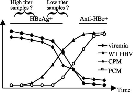
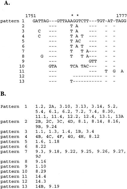
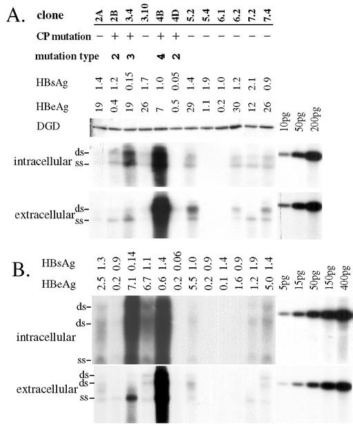
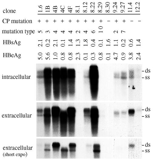
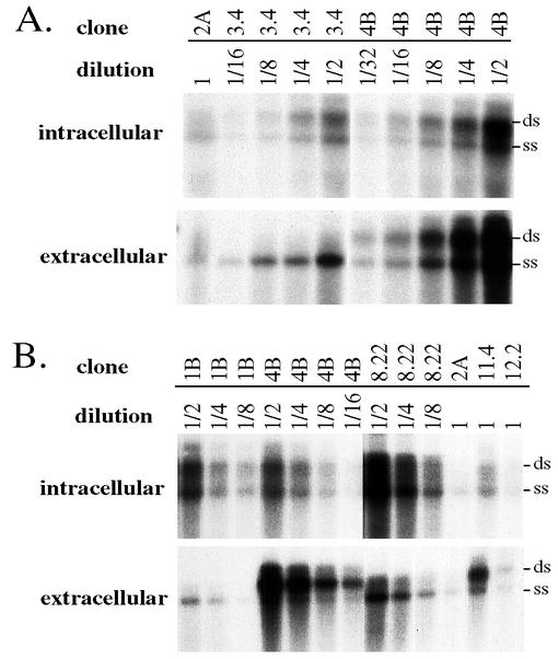
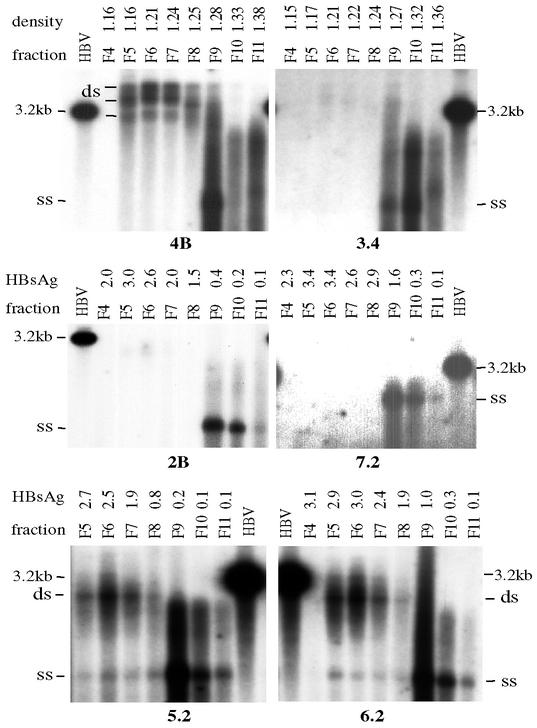
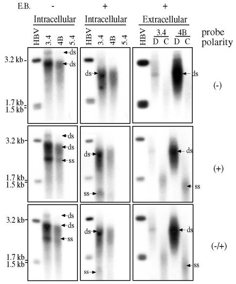
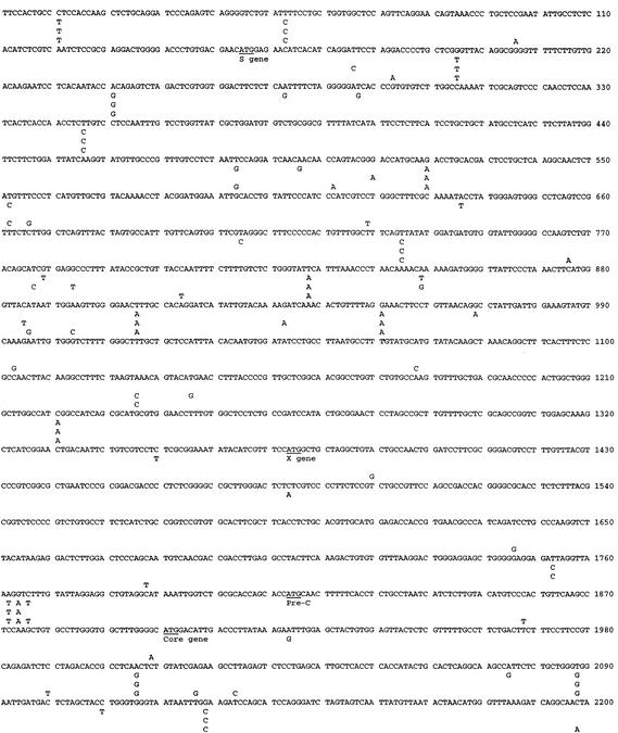
The core promoter mutants of hepatitis B virus (HBV) emerge as the dominant viral population at the late HBeAg and the anti-HBe stages of HBV infection, with the A1762T/G1764A substitutions as the hotspot mutations. The double core promoter mutations were found by many investigators to moderately enhance viral genome replication and reduce hepatitis B e antigen (HBeAg) expression. A much higher replication capacity was reported for a naturally occurring core promoter mutant implicated in the outbreak of fulminant hepatitis, which was caused by the neighboring C1766T/T1768A mutations instead. To systemically study the biological properties of naturally occurring core promoter mutants, we amplified full-length HBV genomes by PCR from sera of HBeAg(+) individuals infected with genotype A. All 12 HBV genomes derived from highly viremic sera (5 x 10(9) to 5.7 x 10(9) copies of viral genome/ml) harbored wild-type core promoter sequence, whereas 37 of 43 clones from low-viremia samples (0.2 x 10(7) to 4.6 x 10(7) copies/ml) were core promoter mutants. Of the 11 wild-type genomes and 14 core promoter mutants analyzed by transfection experiments in human hepatoma cell lines, 6 core promoter mutants but none of the wild-type genomes replicated at high levels. All had 1762/1764 mutations and an additional substitution at position 1753 (T to C), at position 1766 (C to T), or both. Moreover, these HBV clones varied greatly in their ability to secrete enveloped viral particles irrespective of the presence of core promoter mutations. High-replication clones with 1762/1764/1766 or 1753/1762/1764/1766 mutations expressed very low levels of HBeAg, whereas high-replication clones with 1753/1762/1764 triple mutations expressed high levels of HBeAg. Experiments with site-directed mutants revealed that both 1762/1764/1766 and 1753/1762/1764/1766 mutations conferred significantly higher viral replication and lower HBeAg expression than 1762/1764 mutations alone, whereas the 1753/1762/1764 triple mutant displayed only mild reduction in HBeAg expression similar to the 1762/1764 mutant. Thus, core promoter mutations other than those at positions 1762 and 1764 can have major impact on viral DNA replication and HBeAg expression.


Figures

FIG. 1.
FIG. 1.
Diagram of the dynamic change of viral populations during seroconversion from HBeAg to anti-HBe. The seroconversion is accompanied by rapid drop in viremia titer. The wild-type (WT) HBV population declines and eventually disappears as a result of the selective pressure of anti-HBe immunity, whereas the core promoter mutants (CPM) and precore mutants (PCM) arise sequentially to replace the wild-type HBV. We propose that the highly viremic samples used in the present study were at an early HBeAg+ stage of infection, whereas the low-viremia samples were close to seroconversion.
FIG. 2.
FIG. 2.
Core promoter sequences found in HBV clones derived from HBeAg+ serum samples with high- or low-viremia titers. (A) Sequence patterns at 1751 to 1777 of the basic core promoter region. Pattern 1 represents the wild-type sequence. Hyphens indicate a lack of nucleotides at these positions. Patterns 2 to 8 contained only substitutions. Insertions were present in patterns 9 to 11, and deletions occurred in sequences under patterns 12 and 13. The most common substitutions at 1762 and 1764 are marked by asterisks. (B) HBV clones with such divergent sequence patterns. For each clone, the number at the beginning identifies the patient (clone 1.2 was derived from patient 1, whereas clone 2A was from patient 2). It is evident that dominant viral populations from patients 1, 4, and 9 had patterns 3, 4, and 7, respectively.
FIG. 3.
FIG. 3.
Transfection of 12 HBV genomes in Huh7 cells (A) and HepG2 cells (B). Cells grown in 10-cm dishes were transfected with 15 μg of HBV dimer and 15 μg of duck glycine decarboxylase (DGD) cDNA (24) and then harvested 5 days later. From the cell lysates, duck glycine decarboxylase expression was determined by Western blotting, and HBV DNA replication was detected by Southern blot from core particles. From the culture supernatant, HBsAg (shown as the optical density at 490 nm) and HBeAg (shown as counts per minute [103]) were measured by commercial kits after 1:4 and 1:5 dilution of samples, respectively. Extracellular viral particles (both Dane particles and naked core particles) were concentrated by ultracentrifugation through sucrose cushions and analyzed by Southern blotting. Various amounts of 3.2-kb linear HBV DNA were run in parallel as a size marker and for quantification. CP mutation, core promoter mutations; ds, double-stranded viral genome; ss, single-stranded viral genome. Gels were run in the absence of ethidium bromide (A) or in its presence (B). The two “ds” bands in panel B may represent relaxed circular DNA and duplex linear DNA, respectively, whereas the “ds” band in panel A appears to be duplex linear.
FIG. 4.
FIG. 4.
Transfection of 14 HBV genomes in Huh7 cells. Cells were harvested at day 5 posttransfection. For released viral particles, a short exposure is also given to better visualize the bands corresponding to single- and double-stranded viral genomes. ds,: double- stranded viral genome; ss, single-stranded viral genome.
FIG. 5.
FIG. 5.
Quantitative analysis of the relative replication and secretion capacities of different HBV clones. (A) Comparison of clones 2A, 3.4, and 4B. (B) Comparison of clones 1B, 2A, 4B, 8.22, 11.4, and 12.2. Huh7 cells were transfected with various HBV constructs and harvested at day 5 posttransfection. HBV DNA from intracellular core particles and extracellular viral particles were analyzed, with the agarose gels run in the absence of ethidium bromide. The entire samples of 2A, 11.4, and 12.2 were loaded into single wells, while serially diluted samples of 1B, 3.4, 4B, and 8.22 were loaded into separate lanes. ds, double-stranded viral genome; ss, single-stranded viral genome. Please note that clones 3.4 and 1B and, to a lesser extent, clone 8.22, released viral particles with predominantly single-stranded viral genome despite a similar ratio of the two DNA forms inside intracellular core particles.
FIG. 6.
FIG. 6.
CsCl gradient separation of naked core particles from Dane particles. Culture supernatants from 10-cm dishes of Huh7 or HepG2 cells were subjected to ultracentrifugation in 10 to 20% sucrose gradient to pellet the viral particles, which were further spun in CsCl gradient. Fractions of 400 μl were collected, dialyzed, and DNA extracted for Southern blot analysis with an HBV DNA probe (for samples with a high-secretion phenotype, only fractions of DNA samples were used for Southern blotting). The results shown for clones 3.4 and 4B were from an experiment in HepG2 cells, whereas data for the other clones were obtained from transfected Huh7 cells. Except for clone 7.2, gels were run in the presence of ethidium bromide to better separate the single-stranded genome from the double-stranded genome. For clones 3.4 and 4B, the density values of the CsCl fractions were determined. The HBsAg values were measured for CsCl fractions of other samples. Note that clones 3.4, 7.2, and 2B secreted very few Dane particles. HBV, 3.2-kb HBV genome; ss, single-stranded viral genome.
FIG. 7.
FIG. 7.
Detection of intracellular and extracellular HBV DNA by strand-specific probes. Huh7 cells were transfected with HBV clones 3.4, 4B, and 5.4 (as a negative control). DNA was isolated from intracellular core particles, triplicate DNA samples were separated in agarose gels in the presence or absence of ethidium bromide as indicated, and hybridized with one of the three probes. Extracellular viral particles were separated by CsCl gradient, and fractions with densities of Dane particles (D) and core particles (C) were pooled. DNA was separated in an agarose gel in the presence of ethidium bromide and hybridized with one of the three probes. HBV DNA linearized with EcoRI (3.2 kb) and EcoRI plus RsrII (1.7 plus 1.5 kb) were run in parallel as size markers. More DNA samples of clone 3.4 were added than 4B. ds, double stranded; ss, single stranded.
FIG. 8.
FIG. 8.
Comparison of the complete nucleotide sequences of HBV clones 2A (top), 4B (second), 3.4 (third), and 8.22 (bottom). These sequences are available in GenBank under accession numbers AF536524, AF537372, AF537371, and AY152726. The translational start sites for envelope, core, HBx, and polymerase protein are indicated.
FIG. 8.
FIG. 8.
Comparison of the complete nucleotide sequences of HBV clones 2A (top), 4B (second), 3.4 (third), and 8.22 (bottom). These sequences are available in GenBank under accession numbers AF536524, AF537372, AF537371, and AY152726. The translational start sites for envelope, core, HBx, and polymerase protein are indicated.
FIG. 9.
FIG. 9.
Effect of different combinations of core promoter mutations on HBV genome replication and HBeAg expression. (A) Site-directed mutants. Clone 4B contains four point mutations in the core promoter compared with clone 2A. Mu1, mu2a, mu4, and Ex2 are clone 2A-based site-directed mutants containing different mutation combinations. Mutated nucleotides are shown in boldface. (B) Results from one transfection experiment. Huh7 cells grown in 6-cm dishes were transfected with 10 μg of HBV DNA and 2 μg of luciferase plasmid and then harvested 5 days later. The values of luciferase were determined from cell lysate. HBsAg and HBeAg values were determined from culture supernatant. (C) HBeAg expression profiles of the mutants. We used HBeAg/HBsAg ratios to minimize the effect of variation in transfection efficiency. The graph is based on results from seven independent experiments in Huh7 cells. Mu2a and Ex2 displayed highest degree of viral DNA replication and the lowest level of HBeAg expression. Their replication and HBeAg expression phenotypes are close to those of the naturally occurring core promoter mutant, clone 4B.

Similar articles

Cited by

References

    1. Baptista, M., A. Kramvis, and M. Kew. 1999. High prevalence of 1762T 1764A mutations in the basic core promoter of hepatitis B virus isolated from Black Africans with hepatocellular carcinoma compared with asymptomatic carriers. Hepatology 29:946-953. - PubMed
    1. Baumert, T., S. Rogers, K. Hasegawa, and T. Liang. 1996. Two core promoter mutations identified in a hepatitis B virus strain associated with fulminant hepatitis result in enhanced viral replication. J. Clin. Investig. 98:2268-2276. - PMC - PubMed
    1. Baumert, T., A. Marrone, J. Vergalla, and T. Liang. 1998. Naturally occurring mutations define a novel function of the hepatitis B virus core promoter in core protein expression. J. Virol. 72:6785-6795. - PMC - PubMed
    1. Bowyer, S., L. van Staden, M. Kew, and J. Sim. 1997. A unique segment of the hepatitis B virus group A genotype identified in isolates from South Africa. J. Gen. Virol. 78:1719-1729. - PubMed
    1. Buckwold, V., Z. Xu, M. Chen, T. Yen, and J. Ou. 1996. Effects of a naturally occurring mutation in the hepatitis B virus basal core promoter on precore gene expression and viral replication. J. Virol. 70:5845-5851. - PMC - PubMed

Publication types

MeSH terms

Substances

LinkOut - more resources