

Specific inhibition of bovine viral diarrhea virus replicase
- PMID: 12767995
- PMCID: PMC156199
- DOI: 10.1128/jvi.77.12.6753-6760.2003
Specific inhibition of bovine viral diarrhea virus replicase
Abstract
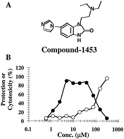
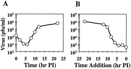
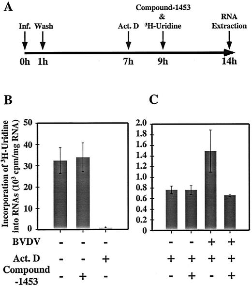
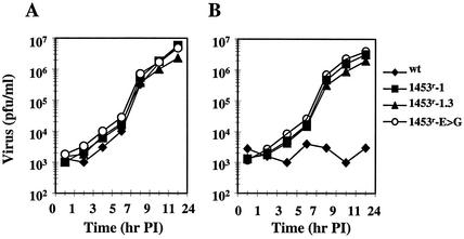
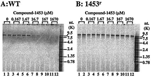
Compound-1453 was identified and characterized as a specific inhibitor of bovine viral diarrhea virus (BVDV). The concentration of compound-1453 which results in 50% protection from virus-induced cytopathic effect is approximately 2.2 microM, with a therapeutic index of 60, and it is not active against a panel of RNA and DNA viruses. A time-of-addition experiment suggested that compound-1453 targets a stage of the viral life cycle after viral entry. To determine the target of compound-1453, resistant virus was generated. Resistant variants grew efficiently in the presence or absence of 33 micro M compound-1453 and exhibited replication efficiency in the presence of compound-1453 approximately 1,000-fold higher than that of the wild-type (wt) virus. Functional mapping and sequence analysis of resistant cDNAs revealed a single amino acid substitution (Glu to Gly) at residue 291 in the NS5B polymerase in all eight independently generated cDNA clones. Recombinant virus containing this single mutation retained the resistance phenotype and a replication efficiency similar to that of the original isolated resistant virus. Since compound-1453 did not inhibit BVDV polymerase activity in vitro (50% inhibitory concentration > 300 microM), we developed a membrane-based assay that consisted of a BVDV RNA replicase complex isolated from virus-infected cells. Compound-1453 inhibited the activity of the wt, but not the drug-resistant, replicase in the membrane assay at concentrations similar to those observed in the viral infection assay. This work presents a novel inhibitor of a viral RNA-dependent RNA replicase.
Figures
Similar articles
-
Substituted 2,6-bis(benzimidazol-2-yl)pyridines: a novel chemical class of pestivirus inhibitors that targets a hot spot for inhibition of pestivirus replication in the RNA-dependent RNA polymerase.Antiviral Res. 2014 Jun;106:71-9. doi: 10.1016/j.antiviral.2014.03.010. Epub 2014 Mar 27. Antiviral Res. 2014. PMID: 24680957
-
Selection of a thiazole urea-resistant variant of bovine viral diarrhoea virus that maps to the RNA-dependent RNA polymerase.Antivir Chem Chemother. 2002 Sep;13(5):315-23. doi: 10.1177/095632020201300507. Antivir Chem Chemother. 2002. PMID: 12630680
-
Highly potent and selective inhibition of bovine viral diarrhea virus replication by γ-carboline derivatives.Antiviral Res. 2010 Dec;88(3):263-8. doi: 10.1016/j.antiviral.2010.09.013. Epub 2010 Sep 24. Antiviral Res. 2010. PMID: 20869990
-
Flaviviridae polymerase and RNA replication.J Viral Hepat. 1999 Jul;6(4):261-70. doi: 10.1046/j.1365-2893.1999.00173.x. J Viral Hepat. 1999. PMID: 10607240 Review.
-
Targeting structural dynamics of the RNA-dependent RNA polymerase for anti-viral strategies.Curr Opin Virol. 2014 Dec;9:194-200. doi: 10.1016/j.coviro.2014.08.006. Epub 2014 Sep 13. Curr Opin Virol. 2014. PMID: 25224392 Review.
Cited by
-
Development of a cell-based high-throughput specificity screen using a hepatitis C virus-bovine viral diarrhea virus dual replicon assay.Antimicrob Agents Chemother. 2005 Apr;49(4):1346-53. doi: 10.1128/AAC.49.4.1346-1353.2005. Antimicrob Agents Chemother. 2005. PMID: 15793110 Free PMC article.
-
Benzimidazole-2-Phenyl-Carboxamides as Dual-Target Inhibitors of BVDV Entry and Replication.Viruses. 2022 Jun 14;14(6):1300. doi: 10.3390/v14061300. Viruses. 2022. PMID: 35746771 Free PMC article.
-
A novel, highly selective inhibitor of pestivirus replication that targets the viral RNA-dependent RNA polymerase.J Virol. 2006 Jan;80(1):149-60. doi: 10.1128/JVI.80.1.149-160.2006. J Virol. 2006. PMID: 16352539 Free PMC article.
-
The imidazopyrrolopyridine analogue AG110 is a novel, highly selective inhibitor of pestiviruses that targets the viral RNA-dependent RNA polymerase at a hot spot for inhibition of viral replication.J Virol. 2007 Oct;81(20):11046-53. doi: 10.1128/JVI.00388-07. Epub 2007 Aug 8. J Virol. 2007. PMID: 17686854 Free PMC article.
-
Chemical genetics strategy identifies an HCV NS5A inhibitor with a potent clinical effect.Nature. 2010 May 6;465(7294):96-100. doi: 10.1038/nature08960. Epub 2010 Apr 21. Nature. 2010. PMID: 20410884 Free PMC article. Clinical Trial.
References
-
- Bartholomeusz, A. I., and P. J. Wright. 1993. Synthesis of dengue virus RNA in vitro: initiation and the involvement of proteins NS3 and NS5. Arch. Virol. 128:111-121. - PubMed
-
- Blumenthal, T., and G. G. Carmichael. 1979. RNA replication: function and structure of QB replicase. Annu. Rev. Biochem. 48:525-548. - PubMed
MeSH terms
Substances
LinkOut - more resources
Full Text Sources
Other Literature Sources
