. 2003 Jun;77(12):6836-44.
doi: 10.1128/jvi.77.12.6836-6844.2003.

Herpes simplex virus glycoprotein K, but not its syncytial allele, inhibits cell-cell fusion mediated by the four fusogenic glycoproteins, gD, gB, gH, and gL

Affiliations

Herpes simplex virus glycoprotein K, but not its syncytial allele, inhibits cell-cell fusion mediated by the four fusogenic glycoproteins, gD, gB, gH, and gL

Elisa Avitabile et al. J Virol. 2003 Jun.

Abstract

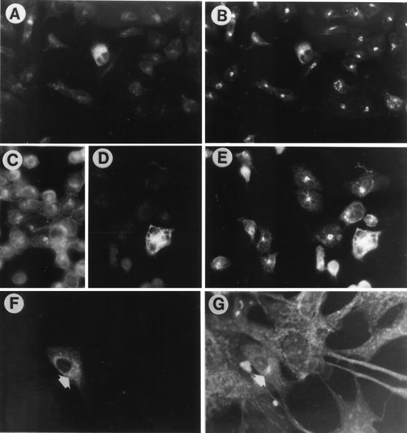
A Myc epitope was inserted at residue 283 of herpes simplex virus type 1 (HSV-1) glycoprotein K (gK), a position previously shown not to interfere with gK activity. The Myc-tagged gK localized predominantly to the endoplasmic reticulum, both in uninfected and in HSV-infected cells. gK, coexpressed with the four HSV fusogenic glycoproteins, gD, gB, gH, and gL, inhibited cell-cell fusion. The effect was partially dose dependent and was observed both in baby hamster kidney (BHK) and in Vero cells, indicating that the antifusion activity of gK may be cell line independent. The antifusion activity of gK did not require viral proteins other than the four fusogenic glycoproteins. A syncytial (syn) allele of gK (syn-gK) carrying the A40V substitution present in HSV-1(MP) did not block fusion to the extent seen with the wild-type (wt) gK, indicating that the syn mutation ablated, at least in part, the antifusogenic activity of wt gK. We conclude that gK is part of the mechanism whereby HSV negatively regulates its own fusion activity. Its effect accounts for the notion that cells infected with wt HSV do not fuse with adjacent, uninfected cells into multinucleated giant cells or syncytia. gK may also function to preclude fusion between virion envelope and the virion-encasing vesicles during virus transport to the extracellular compartment, thus preventing nucleocapsid de-envelopment in the cytoplasm.


Figures

FIG. 1.
FIG. 1.
Radioimmunoprecipitation of Myc-tagged gK. BHK cells were transfected with pwtgK-MTS encoding the Myc-tagged gK or left untransfected. Cells were labeled with a mixture of [35S]methionine and cysteine and harvested 36 h after transfection. Anti-Myc antibody was used to immunoprecipitate gK. The immunoprecipitated proteins were separated by SDS-PAGE and visualized by phosphorimager analysis. Left lane, molecular weight (MW) markers; figures on left, molecular weights of markers, in kilodaltons. The arrow points to gK.
FIG. 2.
FIG. 2.
Pairs of micrographs of 143-tk and Vero cells transfected with pwtgK-MTS and double stained with antibody to Myc (A, C, and E), to calnexin (B and D), or ConA (F). Anti-Myc antibody only stained the transfected cells, whereas the antibody to calnexin and the lectin stained all cells. In panels C and D, transfected cells were superinfected with R7032 at 24 h after transfection and fixed 10 h later. Panels A, C, and E show that anti-Myc staining was reticular and diffused to the cytoplasm, with a pattern indistinguishable from that of calnexin or ConA (arrows), in both uninfected and R7032-infected cells. Panels A to D, 143-tk cells; magnification, ×40. Panels E and F, Vero cells; magnification, ×63.
FIG. 3.
FIG. 3.
Pairs of micrographs of 143-tk and Vero cells transfected with pwtgK-MTS and double stained with antibody to Myc (A, D, and F) and to giantin (B and E) or WGA (G). Anti-Myc antibody only stained the transfected cells, whereas the MAb to giantin and WGA stained all cells. In panels C to E, transfected cells were superinfected with R7032 24 h after transfection and fixed 10 h later. In panel C, cells were stained with anti-gD MAb to show that all cells in the culture were indeed infected. Panels A, D, and F show that anti-Myc staining was reticular and diffused to cytoplasm. This pattern overlaps with that of giantin in infected cells (E) but not in the uninfected cells (B and G). Panels A to E, 143-tk cells; magnification, ×40. Panels F and G, Vero cells; magnification, ×63.
FIG. 4.
FIG. 4.
Micrographs of BHK cells cotransfected with the plasmids encoding gB, gD, gH, and gL and stained with MAbs to gB (A), gD (B), gH (C), and gL (D).
FIG. 5.
FIG. 5.
Digital micrographs of BHK (upper and middle images) or Vero (lower images) cells transfected with the four glycoproteins (gB, gD, gH, and gL) in the absence (left) or presence of wt-gK (middle) or in the presence of syn-gK (right). All cultures were also transfected with a LacZ plasmid. Cells were stained with X-Gal. Middle images show higher magnifications of upper panels. BHK cells received 40 ng (each) of gB, gD, gH, and gL plasmids and 160 ng of gK plasmid. Vero cells received 80 ng (each) of gB, gD, gH, and gL plasmids and 160 ng of gK plasmid.
FIG. 6.
FIG. 6.
Quantification of effect of wt gK and syn-gK on syncytium formation in BHK cells. Digital micrographs of cultures transfected with 20 ng (or 40 ng) (each) of plasmid DNA for the four glycoproteins (gD, gB, gH, and gL), 160 ng of gK plasmid or VP22-Myc plasmid, and 80 ng of LacZ plasmid. Cells were stained with X-Gal 24 or 48 h after transfection. Areas of syncytia were quantified as detailed in Materials and Methods. At least 6 pictures were scored for each sample. The average is expressed in each bar. Thin lines represent standard errors.
FIG. 7.
FIG. 7.
Quantification of effect of wt gK and syn-gK on syncytium formation in Vero cells. Digital micrographs of cultures transfected with 80 ng (each) of gD, gB, gH, and gL, 160 ng of gK plasmid, and 80 ng of LacZ plasmid. Cells were stained with X-Gal 48 h after transfection. Areas of syncytia were quantified as detailed in Materials and Methods. The average is expressed in the each bar. Thin lines represent standard errors.

References

    1. Baines, J. D., and B. Roizman. 1993. The UL10 gene of herpes simplex virus 1 encodes a novel viral glycoprotein, gM, which is present in the virion and in the plasma membrane of infected cells. J. Virol. 67:1441-1452. - PMC - PubMed
    1. Baines, J. D., P. L. Ward, G. Campadelli-Fiume, and B. Roizman. 1991. The UL20 gene of herpes simplex virus 1 encodes a function necessary for viral egress. J. Virol. 65:6414-6424. - PMC - PubMed
    1. Bond, V. C., and S. Person. 1984. Fine structure physical map locations of alterations that affect cell fusion in herpes simplex virus type 1. Virology 132:368-376. - PubMed
    1. Brandimarti, R., T. Huang, B. Roizman, and G. Campadelli-Fiume. 1994. Mapping of herpes simplex virus 1 genes with mutations which overcome host restrictions to infection. Proc. Natl. Acad. Sci. USA 91:5406-5410. - PMC - PubMed
    1. Browne, H., B. Bruun, and T. Minson. 2001. Plasma membrane requirements for cell fusion induced by herpes simplex virus type 1 glycoproteins gB, gD, gH and gL. J. Gen. Virol. 82:1419-1422. - PubMed

Publication types

Substances

LinkOut - more resources