. 2003 Jun;77(12):6845-54.
doi: 10.1128/jvi.77.12.6845-6854.2003.

Follicular dendritic cell dedifferentiation by treatment with an inhibitor of the lymphotoxin pathway dramatically reduces scrapie susceptibility

Affiliations

Follicular dendritic cell dedifferentiation by treatment with an inhibitor of the lymphotoxin pathway dramatically reduces scrapie susceptibility

Neil A Mabbott et al. J Virol. 2003 Jun.

Abstract

Transmissible spongiform encephalopathies (TSEs) may be acquired peripherally, in which case infectivity usually accumulates in lymphoid tissues before dissemination to the nervous system. Studies of mouse scrapie models have shown that mature follicular dendritic cells (FDCs), expressing the host prion protein (PrP(c)), are critical for replication of infection in lymphoid tissues and subsequent neuroinvasion. Since FDCs require lymphotoxin signals from B lymphocytes to maintain their differentiated state, blockade of this stimulation with a lymphotoxin beta receptor-immunoglobulin fusion protein (LT beta R-Ig) leads to their temporary dedifferentiation. Here, a single treatment with LT beta R-Ig before intraperitoneal scrapie inoculation blocked the early accumulation of infectivity and disease-specific PrP (PrP(Sc)) within the spleen and substantially reduced disease susceptibility. These effects coincided with an absence of FDCs in the spleen for ca. 28 days after treatment. Although the period of FDC dedifferentiation was extended to at least 49 days by consecutive LT beta R-Ig treatments, this had little added protective benefit after injection with a moderate dose of scrapie. We also demonstrate that mature FDCs are critical for the transmission of scrapie from the gastrointestinal tract. Treatment with LT beta R-Ig before oral scrapie inoculation blocked PrP(Sc) accumulation in Peyer's patches and mesenteric lymph nodes and prevented neuroinvasion. However, treatment 14 days after oral inoculation did not affect survival time or susceptibility, suggesting that infectivity may have already spread to the peripheral nervous system. Although manipulation of FDCs may offer a potential approach for early intervention in peripherally acquired TSEs, these data suggest that the duration of the treatment window may vary widely depending on the route of exposure.


Figures
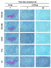
FIG. 1.
FIG. 1.
Effect of LTβR-Ig treatment on FDC status in spleens of uninfected mice. Tissues were taken on the days indicated postinjection with hu-Ig (control) or LTβR-Ig, and adjacent frozen sections stained with FDC-M1 (top row; red) and FDC-M2 (second row; red) monoclonal antiserum to detect FDCs, 8C12 monoclonal antiserum to detect CD35 (third row; red), and monoclonal antiserum specific for complement components C1q (fourth row; red) and C3 (lower row; red). Expression of FDC-M1, FDC-M2, CD35, and associated accumulations of complement components C1q and C3 were undetectable in the spleen after treatment with LTβR-Ig. Original magnification, ×400.
FIG. 2.
FIG. 2.
Treatment with LTβR-Ig prior to i.p. scrapie inoculation blocks the early accumulation of PrPSc and infectivity in the spleen. Mice were treated with LTβR-Ig or hu-Ig (as a control) 3 days before i.p. scrapie inoculation, and tissues were assayed 70 days after inoculation (a) or at the terminal stage of disease (b). Immunoblots show the accumulation of detergent insoluble, relatively proteinase K (PK)-resistant PrPSc. Treatment of tissue in the presence (+) or absence (−) of proteinase K before electrophoresis is indicated. After proteinase K treatment, a typical three-band pattern was observed between molecular mass values of 20 and 30 kDa, representing unglycosylated, monoglycosylated, and diglycosylated isomers of PrP (in order of increasing molecular mass). PrP was detected by using the PrP-specific monoclonal antiserum 8H4. Lane M contained molecular mass markers. The scrapie infectivity titer is expressed as the log i.c. ID50 units/g. (a) A total of 1 of 12 assay mice developed clinical scrapie within 500 days of inoculation; (b) 2 of 12 assay mice developed clinical scrapie within 500 days of inoculation. dpi, day postinoculation on which the tissues were analyzed; pos., mice that developed clinical signs of scrapie; neg., mice that were free of the clinical signs of scrapie.
FIG. 3.
FIG. 3.
Effect of multiple hu-Ig or LTβR-Ig treatments on FDC status in spleen. Mice were given a single i.p. injection of 100 μg of LTβR-Ig (or 100 μg of hu-Ig as a control) 3 days before i.p. inoculation with scrapie. After scrapie challenge, mice were then given one or two subsequent doses of 100 μg of LTβR-Ig or 100 μg of hu-Ig at 21-day intervals (days 21 and 42 postinoculation with scrapie). Spleens were taken from two mice from each group 7 days after the last treatment (day 7, day 28, or day 49 after inoculation with scrapie for mice given one, two or three consecutive treatments, respectively). Adjacent frozen sections were stained with FDC-M2 monoclonal antiserum to detect FDCs (top row; red), 8C12 monoclonal antiserum to detect CD35 (second row; red), and monoclonal antisera specific for complement components C1q (third row; red) and C3 (fourth row; red). All sections were counterstained with hematoxylin (blue). After treatment of mice with three consecutive doses of LTβR-Ig, the expression of FDC-M2, CD35, and associated accumulations of complement components C1q and C3 remained undetectable for up to at least 49 days after the first treatment. Original magnification, ×200.
FIG. 4.
FIG. 4.
Effect of LTβR-Ig treatment on FDC status in Peyer's patches and MLNs. Tissues were obtained 3 days postinjection with hu-Ig (control; upper panels) or LTβR-Ig (lower panels), and sections were stained with FDC-M2 monoclonal antiserum (red) to detect FDCs. All sections were counterstained with hematoxylin (blue). Mature FDC-M2-expressing FDCs were undetectable in Peyer's patches and MLNs after treatment with LTβR-Ig. Original magnification, ×200.
FIG. 5.
FIG. 5.
Immunohistological analysis of brain tissue from mice treated with hu-Ig or LTβR-Ig 3 days before (−3) or 14 days after (+14) oral inoculation with scrapie. Large disease-specific PrP accumulations (upper row; brown), reactive astrocytes expressing high levels of GFAP (middle row; red), and spongiform pathology (H&E; lower row) were detected in the hippocampi of all mice showing clinical signs of scrapie. In contrast, in the brains of mice treated with LTβR-Ig 3 days before inoculation (LTβR-Ig −3), no evidence of PrP accumulation, reactive astrocytes, or spongiform pathology was detected 518 days after inoculation. All sections were counterstained with hematoxylin (blue). dpi, day postinoculation on which the tissues were analyzed; pos., mice that developed clinical signs of scrapie; neg., mice that were free of the clinical signs of scrapie. Original magnification, ×100.
FIG. 6.
FIG. 6.
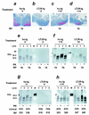
Treatment with LTβR-Ig prior to oral scrapie inoculation blocks the accumulation of disease-specific PrP in Peyer's patches, MLNs, and spleens. Mice were treated with LTβR-Ig or hu-Ig as a control 3 days before (a, b, e, and g; −3) or 14 days after (c, d, f, and h; +14) oral inoculation with scrapie. Disease-specific PrP accumulations were determined in Peyer's patches (a to d) and MLNs (e and f) assayed 70 days after inoculation and in the spleen (g and h) at the terminal stage of disease. In panels a to d, PrP accumulations in Peyer's patches were detected on paraffin-embedded sections with the PrP-specific polyclonal antiserum 1B3 (PrP, red). Sections were counterstained with hematoxylin (blue). Original magnification, ×100. In panels e to h, immunoblots show the accumulation of detergent-insoluble, relatively proteinase K (PK)-resistant PrPSc in the MLNs (e and f) and spleens (g and h) of treated mice. Treatment of the tissue in the presence (+) or absence (−) of proteinase K before electrophoresis is indicated. PrP was detected by using the PrP-specific monoclonal antiserum 8H4. Lane M contained molecular mass markers. dpi, day postinoculation on which the tissues were analyzed; pos., mice that developed clinical signs of scrapie; neg., mice that were free of the clinical signs of scrapie.

References

    1. Andreoletti, O., P. Berthon, D. Marc, P. Sarradin, J. Grosclaude, L. van Keulen, F. Schelcher, J.-M. Elsen, and F. Lantier. 2000. Early accumulation of PrPSc in gut-associated lymphoid and nervous tissues of susceptible sheep from a Romanov flock with natural scrapie. J. Gen. Virol. 81:3115-3126. - PubMed
    1. Aucouturier, P., F. Geissmann, D. Damotte, G. P. Saborio, H. C. Meeker, R. Kascsak, R. Kascsak, R. I. Carp, and T. Wisniewski. 2001. Infected splenic dendritic cells are sufficient for prion transmission to the CNS in mouse scrapie. J. Clin. Investig. 108:703-708. - PMC - PubMed
    1. Beekes, M., and P. A. McBride. 2000. Early accumulation of pathological PrP in the enteric nervous system and gut-associated lymphoid tissue of hamsters orally infected with scrapie. Neurosci. Lett. 278:181-184. - PubMed
    1. Beringue, V., M. Demoy, C. I. Lasmezas, B. Gouritin, C. Weingarten, J.-P. Deslys, J.-P. Adreux, P. Couvreur, and D. Dormont. 2000. Role of spleen macrophages in the clearance of scrapie agent early in pathogenesis. J. Pathol. 190:495-502. - PubMed
    1. Bolton, D. C., M. P. McKinley, and S. B. Prusiner. 1982. Identification of a protein that purifies with the scrapie prion. Science 218:1309-1311. - PubMed

Publication types

MeSH terms