. 2003 Jun;77(12):6931-45.
doi: 10.1128/jvi.77.12.6931-6945.2003.

Targeting of the human immunodeficiency virus type 1 envelope to the trans-Golgi network through binding to TIP47 is required for env incorporation into virions and infectivity

Affiliations

Targeting of the human immunodeficiency virus type 1 envelope to the trans-Golgi network through binding to TIP47 is required for env incorporation into virions and infectivity

Guillaume Blot et al. J Virol. 2003 Jun.

Abstract

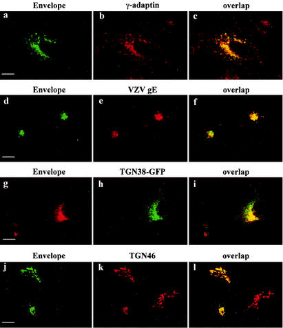
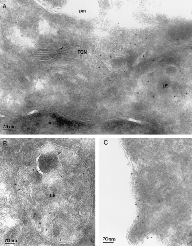
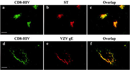
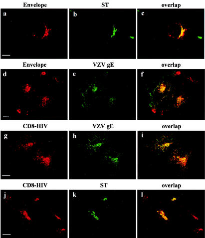
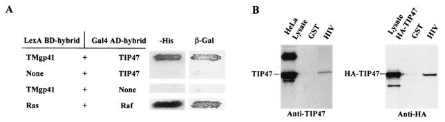
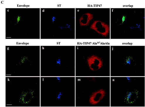
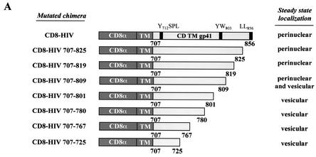
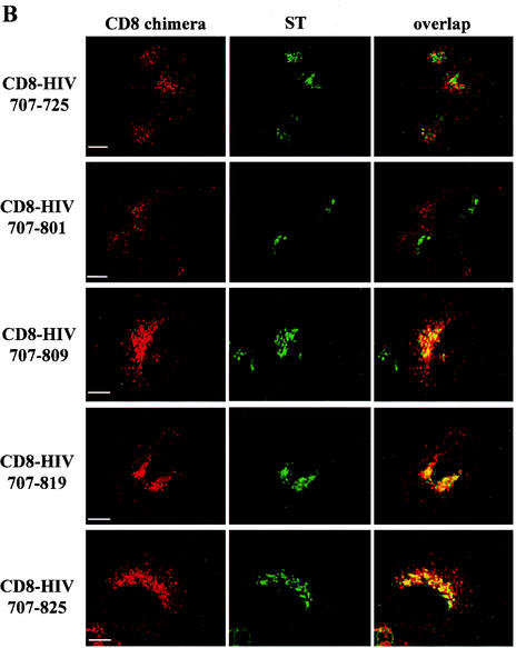
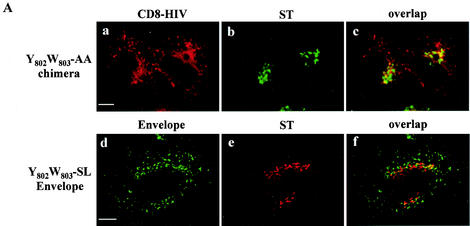
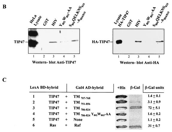
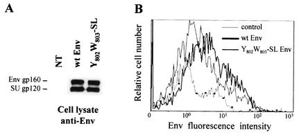
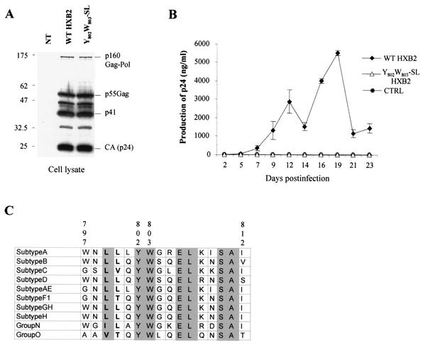
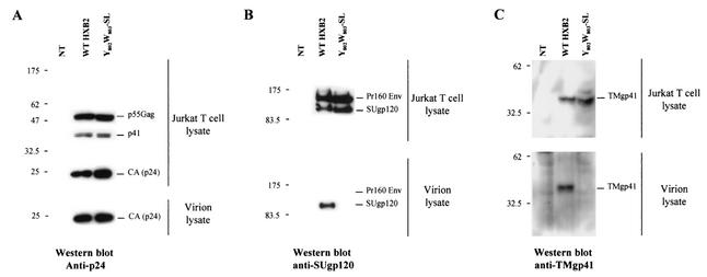
Here, we report that human immunodeficiency virus type 1 (HIV-1) Env glycoprotein is located mainly in the trans-Golgi network (TGN) due to determinants present in the cytoplasmic domain of the transmembrane gp41 glycoprotein (TMgp41). Internalization assays demonstrated that Env present at the cell surface returns to the TGN. We found that the cytoplasmic domain of TMgp41 binds to TIP47, a protein required for the transport of mannose-6-phosphate receptors from endosomes to the TGN. Overexpression of a mutant of TIP47 affected the transport of Env from endosomes to the TGN. Retrograde transport of Env to the TGN requires a Y(802)W(803) diaromatic motif present in the TMgp41 cytoplasmic domain. Mutation of this motif abolished both targeting to the TGN as well as interaction with TIP47. These data support the view that binding of TIP47 to HIV-1 Env facilitates its delivery to the TGN. Lastly, we show that virus mutated in the Y(802)W(803) motif is poorly infectious and presents a defect in Env incorporation, supporting a model in which retrograde transport of Env is implicated in the optimization of fully infectious HIV-1 production.


Figures

FIG. 1.
FIG. 1.
Localization of HIV-1 Env glycoprotein by confocal microscopy. HeLa cells were transfected with pcEnv-wt (a to c) or cotransfected with pcEnv-wt and pSFFI-gE (d to f), or pcEnv-wt and ΔpMEP4-TGN38EGFP (g to i). P4-2 HeLa cells were infected with HXB2 HIV-1 (j to l). The transfected and infected cells were fixed and processed for immunofluorescence with the b12 anti-Env MAb (a, d, g, and j) and either the anti-γ-adaptin MAb (b) or antibodies against VZV gE (e) or TGN46 (k). Cells were treated with 50 μg of cycloheximide/ml for 3 h before immunofluorescence. Colocalization was examined by confocal microscopy. A series of optical sections at 0.5-μm intervals was recorded. A representative medial section is shown. Scale bar, 20 μm.
FIG. 2.
FIG. 2.
Localization of Env by immunoelectron microscopy. Ultrathin cryosections of HeLa cells expressing Env immunogold-labeled with anti-Env 2G12 (10-nm-diameter gold particles). (A) At the steady state, Env was detected in the Golgi cisternae and tubulo-vesicular structures of the TGN (TGN). n, nucleus. On the right, a multivesicular LE was labeled with 2G12 MAb. (B) Multivesicular LE labeled with 2G12 MAb. (C) At the steady state, immunogold labeling of Env appears scattered along the plasma membrane (pm).
FIG. 3.
FIG. 3.
Localization of CD8-HIV chimeric molecules by confocal microscopy. HeLa cells were cotransfected with pCD8-HIV with either pSRα-STVSVG (a to c) or pSFFI-gE (d to f). The transfected cells were fixed and processed for immunofluorescence with the CD8-Leu2A MAb (a and d) and antibodies against ST (b) or VZV gE (e). Before fixation, all the cells were treated with 50 μg of cycloheximide/ml for 3 h. Colocalization was examined by confocal microscopy as described in the legend to Fig. 1. Scale bar, 20 μm.
FIG. 4.
FIG. 4.
Internalization of HIV-1 Env glycoprotein and CD8-HIV chimeric molecules. HeLa cells expressing either Env and ST (a and b), CD8-HIV chimera and VZV gE (g and h), or CD8-HIV chimera and ST (j and k) were incubated at 0°C with the b12 anti-Env (a) or Leu2A anti-CD8 (g and j) MAbs for 1 h and then returned to 37°C for 1 h. The cells were fixed, permeabilized, and labeled with antibodies against either ST (b and k) or VZV gE (h). For uptake experiments (d), HeLa cells expressing Env and VZV gE (d to f) were incubated for 1 h at 37°C with the b12 anti-Env (d) and anti-VZV gE (e) antibodies. The cells were fixed, permeabilized, and stained with the appropriate secondary antibodies (d to f). Colocalization was examined by confocal microscopy as described in the legend to Fig. 1. Scale bar, 20 μm.
FIG. 5.
FIG. 5.
Binding of the cytosolic tail of Env to TIP47 (A) Interaction of the cytosolic tail of Env with TIP47 in yeast two-hybrid assay. The yeast reporter strain L40 expressing the indicated hybrid protein pairs was analyzed for histidine auxotrophy (−His) and β-Gal expression. The full-length cytosolic tail of TMgp41 was fused to the LexA BD. The TIP47 ORF was fused to the Gal4 activation domain (Gal4AD). Ras and Raf proteins, which bind to each other efficiently, were used as positive controls. (B) Binding of the cytosolic tail of Env to TIP47 in HeLa cell lysate binding assay. HeLa cell lysates expressing endogenous TIP47 (left panel) or HA-TIP47 (right panel) were incubated with equal amounts of purified GST or GST fused to the cytoplasmic tail of HIV-1 TMgp41 (GST-HIV). Bound materials were analyzed by SDS-PAGE, and TIP47 binding was analyzed by Western blotting with either anti-TIP47 (left panel) or anti-HA MAb (right panel). Crude lysates corresponding to 2 × 10⁵ cells were also run as a control to detect endogenous TIP47 (lane HeLa lysate in left panel) or HA-TIP47 (lane lysate HA-TIP47 in right panel). (C) Transdominant effect of TIP47Ala167AlaAla on Env retrograde transport. HeLa cells expressing Env, ST, and HA-TIP47 (upper panel) or HA-TIP47Ala167AlaAla mutant (lower panels) were incubated at 0°C with the b12 anti-Env MAb for 1 h and then returned to 37°C for 1 h. The cells were fixed, permeabilized, and labeled with antibodies directed against ST and the HA epitope. Colocalization was examined by confocal microscopy as described in the legend to Fig. 1. Overlaps between Env and ST labeling were presented in panels f, j, and n. Scale bar, 20 μm.
FIG. 5.
FIG. 5.
Binding of the cytosolic tail of Env to TIP47 (A) Interaction of the cytosolic tail of Env with TIP47 in yeast two-hybrid assay. The yeast reporter strain L40 expressing the indicated hybrid protein pairs was analyzed for histidine auxotrophy (−His) and β-Gal expression. The full-length cytosolic tail of TMgp41 was fused to the LexA BD. The TIP47 ORF was fused to the Gal4 activation domain (Gal4AD). Ras and Raf proteins, which bind to each other efficiently, were used as positive controls. (B) Binding of the cytosolic tail of Env to TIP47 in HeLa cell lysate binding assay. HeLa cell lysates expressing endogenous TIP47 (left panel) or HA-TIP47 (right panel) were incubated with equal amounts of purified GST or GST fused to the cytoplasmic tail of HIV-1 TMgp41 (GST-HIV). Bound materials were analyzed by SDS-PAGE, and TIP47 binding was analyzed by Western blotting with either anti-TIP47 (left panel) or anti-HA MAb (right panel). Crude lysates corresponding to 2 × 10⁵ cells were also run as a control to detect endogenous TIP47 (lane HeLa lysate in left panel) or HA-TIP47 (lane lysate HA-TIP47 in right panel). (C) Transdominant effect of TIP47Ala167AlaAla on Env retrograde transport. HeLa cells expressing Env, ST, and HA-TIP47 (upper panel) or HA-TIP47Ala167AlaAla mutant (lower panels) were incubated at 0°C with the b12 anti-Env MAb for 1 h and then returned to 37°C for 1 h. The cells were fixed, permeabilized, and labeled with antibodies directed against ST and the HA epitope. Colocalization was examined by confocal microscopy as described in the legend to Fig. 1. Overlaps between Env and ST labeling were presented in panels f, j, and n. Scale bar, 20 μm.
FIG. 6.
FIG. 6.
Representation of the cytoplasmic tail of wt HIV-1 Env and of the mutants used in this study. (A) The external domain and the anchor peptide (TM) of the human CD8α chain antigen are indicated. Numbering corresponds to the position of the beginning and end of the HIV-1 TMgp41 cytoplasmic domain (CD TMgp41). The steady-state locations of the different mutants are judged by immunofluorescence analysis. (B) Effect of deletion of the TMgp41 cytoplasmic domain on return of the CD8-HIV chimeric molecule to the TGN. HeLa cells were transfected with pSRα-STVSVG and either pCD8-HIV707-725, pCD8-HIV707-801, pCD8-HIV707-809, pCD8-HIV707-819, or pCD8-HIV 707-825 vector. The transfected cells were incubated at 0°C with the CD8-Leu2A MAb for 1 h and then returned to 37°C for 1 h. The cells were fixed, permeabilized, and labeled with anti-ST antibodies. Colocalization was examined by confocal microscopy as described in the legend to Fig. 1. Scale bar, 20 μm.
FIG. 6.
FIG. 6.
Representation of the cytoplasmic tail of wt HIV-1 Env and of the mutants used in this study. (A) The external domain and the anchor peptide (TM) of the human CD8α chain antigen are indicated. Numbering corresponds to the position of the beginning and end of the HIV-1 TMgp41 cytoplasmic domain (CD TMgp41). The steady-state locations of the different mutants are judged by immunofluorescence analysis. (B) Effect of deletion of the TMgp41 cytoplasmic domain on return of the CD8-HIV chimeric molecule to the TGN. HeLa cells were transfected with pSRα-STVSVG and either pCD8-HIV707-725, pCD8-HIV707-801, pCD8-HIV707-809, pCD8-HIV707-819, or pCD8-HIV 707-825 vector. The transfected cells were incubated at 0°C with the CD8-Leu2A MAb for 1 h and then returned to 37°C for 1 h. The cells were fixed, permeabilized, and labeled with anti-ST antibodies. Colocalization was examined by confocal microscopy as described in the legend to Fig. 1. Scale bar, 20 μm.
FIG.7.
FIG.7.
Effect of mutations in the Y802W803 aromatic doublet on internalization of CD8-HIV chimera and Env glycoprotein. (A) HeLa cells expressing either mutated chimera CD8-HIV Y802W803-AA and ST (a to c) or Y802W803-SL-mutated Env and ST (d to f) were incubated at 0°C with the anti-CD8 Leu2A (a) or the b12 anti-Env (d) MAbs for 1 h and then returned to 37°C for 1 h. The cells were fixed, permeabilized, and labeled with anti-ST antibodies (b and e). Colocalization was examined by confocal microscopy (f). (B) Binding of the mutated cytosolic tail of Env to TIP47. HeLa cell lysates expressing endogenous TIP47 (left panel) or HA-TIP47 (right panel) were incubated with equal amounts of purified GST, GST-HIV, Y802W803-AA, and S804QELKNS810-A804/810 mutated GST-HIV. Bound materials were separated by SDS-PAGE, and TIP47 binding was analyzed by Western blotting with anti-TIP47 (left panel) or with anti-HA MAb (right panel). Crude lysates corresponding to 2 × 10⁵ cells were also run as a control to detect TIP47 or HA-TIP47. (C) Analysis of the ability of the TMgp41 deletion mutant to interact with TIP47 in the two-hybrid assay. The yeast reporter strain L40 expressing the indicated hybrid protein pairs was analyzed for β-Gal expression. The TMgp41 deletion mutants indicated were fused to the Gal4 activation domain (Gal4AD). The TIP47 ORF was fused to the LexA BD. Ras and Raf proteins, which bind to each other efficiently, were used as positive controls.
FIG.7.
FIG.7.
Effect of mutations in the Y802W803 aromatic doublet on internalization of CD8-HIV chimera and Env glycoprotein. (A) HeLa cells expressing either mutated chimera CD8-HIV Y802W803-AA and ST (a to c) or Y802W803-SL-mutated Env and ST (d to f) were incubated at 0°C with the anti-CD8 Leu2A (a) or the b12 anti-Env (d) MAbs for 1 h and then returned to 37°C for 1 h. The cells were fixed, permeabilized, and labeled with anti-ST antibodies (b and e). Colocalization was examined by confocal microscopy (f). (B) Binding of the mutated cytosolic tail of Env to TIP47. HeLa cell lysates expressing endogenous TIP47 (left panel) or HA-TIP47 (right panel) were incubated with equal amounts of purified GST, GST-HIV, Y802W803-AA, and S804QELKNS810-A804/810 mutated GST-HIV. Bound materials were separated by SDS-PAGE, and TIP47 binding was analyzed by Western blotting with anti-TIP47 (left panel) or with anti-HA MAb (right panel). Crude lysates corresponding to 2 × 10⁵ cells were also run as a control to detect TIP47 or HA-TIP47. (C) Analysis of the ability of the TMgp41 deletion mutant to interact with TIP47 in the two-hybrid assay. The yeast reporter strain L40 expressing the indicated hybrid protein pairs was analyzed for β-Gal expression. The TMgp41 deletion mutants indicated were fused to the Gal4 activation domain (Gal4AD). The TIP47 ORF was fused to the LexA BD. Ras and Raf proteins, which bind to each other efficiently, were used as positive controls.
FIG. 8.
FIG. 8.
Expression of Env mutated in Y802W803 at the cell surface. (A) Identical quantities of lysates from transfected cells were analyzed by Western blotting with anti-Env MAb. NT, untransfected control cells. (B) 293T cells (2 × 10⁶) were cotransfected by the calcium phosphate method with 8 μg of wt (thick line) or Y802W803-SL (thin line) Env expression vector and 0.5 μg of pRSV-GFP vector. The amount of Env at the surface of GFP+ cells was analyzed by flow cytometry 48 h later. Dotted line, cells transfected with the pRSV-GFP vector alone, as the negative control (ctrl). Data are representative of three independent experiments.
FIG. 9.
FIG. 9.
Replication in Jurkat T cells of wt and Y802W803-SL mutant HIV-1 virus. (A) Expression of Gag (p160Gag-Pol, p41, p55Gag, or CAp24) proteins of wt and Y802W803-SL mutant HXB2 HIV-1 provirus in transfected 293T cells was verified by Western blot analysis with anti-p24 antibodies. NT, untransfected cells. (B) Replication kinetics. The Jurkat T-cell line was infected in parallel with a low inoculum of wt and Y802W803-SL mutant HXB2 HIV-1 (50 ng of p24/106 cells). Cells were split every 2 or 3 days; culture supernatant samples were collected at each point for p24 antigen quantification. Values are the averages of 3 independent experiments ± standard deviations. CTRL, noninfected cells. (C) Sequence comparison for residues 797 to 812 of the Env cytosolic domain of the different HIV-1 subgroups. The amino acid sequences of representative isolates of different clades (21) (A, U455; B, HXB2R; C, ETH2220; D, ELI; AE, 92TH011A; F1, 93BR020.1; GH, AG-VI525A2; H, 90CR056; O, ANT70) of the HIV-1 groups M, N, and O were aligned. YW is shown in boldface.
FIG. 10.
FIG. 10.
Analysis of wt and Y802W803-SL mutant Env incorporation into virions. Analysis of Gag (p160Gag-Pol, p41, p55Gag, or CAp24) and Env (Pr160 env, SUgp120, or TMgp41) content was analyzed by Western blotting. Cell and virion lysates were prepared from Jurkat T cells infected with wt and Y802W803-SL virus stocks pseudotyped with VSV-G. Samples were transferred to polyvinylidene difluoride membranes and blotted with either an anti-p24 (CA) (A), an anti-SUgp120 (B), or an anti-TMgp41 (C) MAb (Materials and Methods). These results are representative of duplicate experiments. NT, untransfected cells.

References

    1. Alconada, A., U. Bauer, and B. Hoflack. 1996. A tyrosine-based motif and a casein kinase II phosphorylation site regulate the intracellular trafficking of the varicella-zoster virus glycoprotein I, a protein localized in the trans-Golgi network. EMBO J. 15:6096-6110. - PMC - PubMed
    1. Barbero, P., E. Buell, S. Zulley, and S. R. Pfeffer. 2001. Tip47 is not a component of lipid droplets. J. Biol. Chem. 276:24348-24351. - PubMed
    1. Berlioz-Torrent, C., B. L. Shacklett, L. Erdtmann, L. Delamarre, I. Bouchaert, P. Sonigo, M. C. Dokhelar, and R. Benarous. 1999. Interactions of the cytoplasmic domains of human and simian retroviral transmembrane proteins with components of the clathrin adaptor complexes modulate intracellular and cell surface expression of envelope glycoproteins. J. Virol. 73:1350-1361. - PMC - PubMed
    1. Blom, J., C. Nielsen, and J. M. Rhodes. 1993. An ultrastructural study of HIV-infected human dendritic cells and monocytes/macrophages. APMIS 101:672-680. - PubMed
    1. Boge, M., S. Wyss, J. S. Bonifacino, and M. Thali. 1998. A membrane-proximal tyrosine-based signal mediates internalization of the HIV-1 envelope glycoprotein via interaction with the AP-2 clathrin adaptor. J. Biol. Chem. 273:15773-15778. - PubMed

Publication types

MeSH terms

LinkOut - more resources