. 2003 Jun;77(12):7007-16.
doi: 10.1128/jvi.77.12.7007-7016.2003.

Human cytomegalovirus infection inhibits tumor necrosis factor alpha (TNF-alpha) signaling by targeting the 55-kilodalton TNF-alpha receptor

Affiliations

Human cytomegalovirus infection inhibits tumor necrosis factor alpha (TNF-alpha) signaling by targeting the 55-kilodalton TNF-alpha receptor

J Baillie et al. J Virol. 2003 Jun.

Abstract

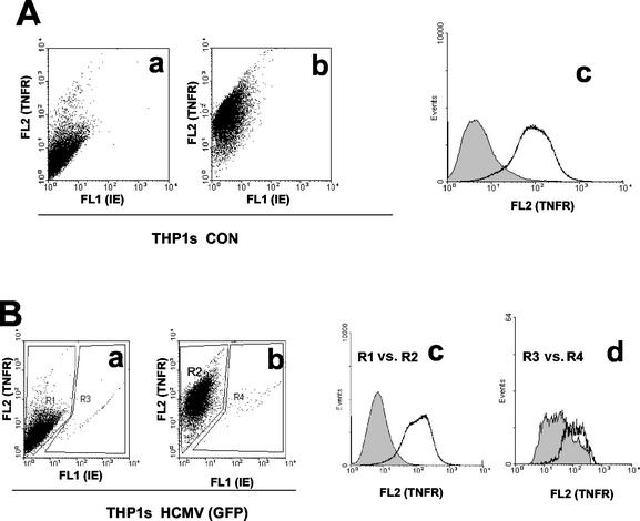
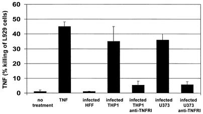
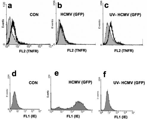
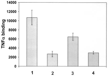
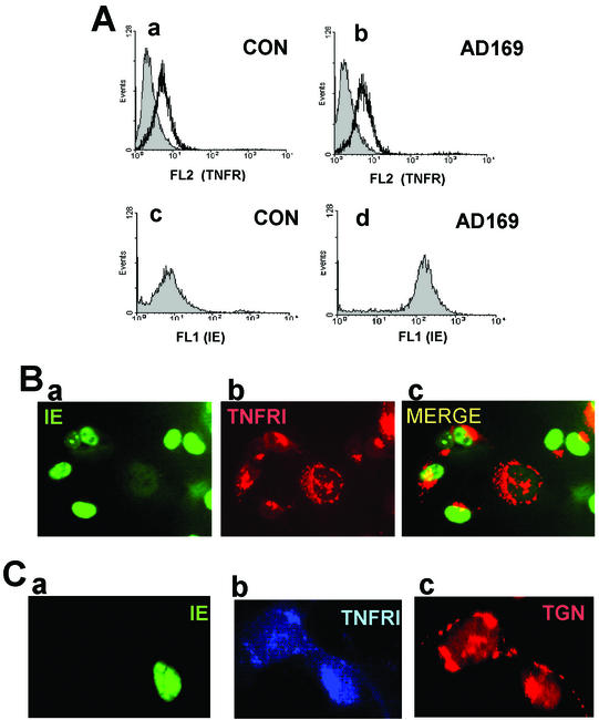
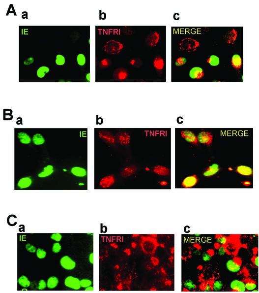
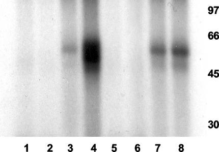
Infection with human cytomegalovirus (HCMV) results in complex interactions between viral and cellular factors which perturb many cellular functions. HCMV is known to target the cell cycle, cellular transcription, and immunoregulation, and it is believed that this optimizes the cellular environment for viral DNA replication during productive infection or during carriage in the latently infected host. Here, we show that HCMV infection also prevents external signaling to the cell by disrupting the function of TNFRI, the 55-kDa receptor for tumor necrosis factor alpha (TNF-alpha), one of the receptors for a potent cytokine involved in eliciting a wide spectrum of cellular responses, including antiviral responses. HCMV infection of fully permissive differentiated monocytic cell lines and U373 cells resulted in a reduction in cell surface expression of TNFRI. The reduction appeared to be due to relocalization of TNFRI from the cell surface and was reflected in the elimination of TNF-alpha-induced Jun kinase activity. Analysis of specific phases of infection suggested that viral early gene products were responsible for this relocalization. However, a mutant HCMV in which all viral gene products known to be involved in down-regulation of major histocompatibility complex (MHC) class I were deleted still resulted in relocalization of TNFRI. Consequently, TNFRI relocalization by HCMV appears to be mediated by a novel viral early function not involved in down-regulation of cell surface MHC class I expression. We suggest that upon infection, HCMV isolates the cell from host-mediated signals, forcing the cell to respond only to virus-specific signals which optimize the cell for virus production and effect proviral responses from bystander cells.


Figures

FIG. 1.
FIG. 1.
HCMV decreases cell surface TNFRI expression in THP1 cells. (A) Uninfected THP1 cells (CON) were stained with a PE-conjugated control antibody (a) or anti-TNFRI antibody (b) as detailed in Materials and Methods. Cells were analyzed by FACS for TNFRI (FL2) and GFP-IE (FL1) staining. The data from the dot plots shown in panels a and b are also presented as a histogram plot (c), with control antibody staining shown by the shaded area and anti-TNFRI antibody staining shown by the white area. (B) THP1 cells infected overnight with GFP-tagged HCMV (5 PFU/cell) were stained with a PE-conjugated control antibody (a) or anti-TNFRI antibody (b) as detailed in Materials and Methods. Cells were analyzed by FACS for TNFRI (FL2) and GFP-IE (FL1) staining. The data from the dot plots shown in panels a and b are also presented as a histogram plot where the specific staining for TNFRI in the uninfected cells in the population is shown as R1 versus R2 (c), and the specific staining for TNFRI in the infected cells in the population is shown as R3 versus R4 (d). In both these histogram plots, control antibody staining is shown by the shaded area, and anti-TNFRI antibody staining is shown by the white area.
FIG. 2.
FIG. 2.
U373 cells and monocytic cells release TNF-α upon infection with HCMV. Using an L929 bioassay, supernatants from uninfected and infected U373, HFF, and THP1 cells were assayed for release of TNF-α. Supernatants were also treated with anti-TNFRI antibody to ensure that the killing observed was TNF-α specific. L929 cells were also treated with purified TNF-α (20 ng/ml) alone. In all cases, values were corrected for any killing observed with control medium.
FIG. 3.
FIG. 3.
HCMV down-regulates cell surface TNFRI expression on U373 cells. Uninfected control (CON) U373 cells (a and d), U373 cells infected overnight with GFP-tagged HCMV (5 PFU/cell) (b and e), or U373 cells infected with UV-inactivated GFP-tagged HCMV (c and f) were stained with a PE-conjugated anti-TNFRI antibody (white area) or a PE-conjugated, isotype-matched control antibody (shaded area) and analyzed by FACS for TNFRI staining (FL2) or GFP-IE staining (FL1).
FIG. 4.
FIG. 4.
HCMV deletions devoid of all MHC class I down-regulating genes still down-regulateTNFRI. (A) Uninfected control (CON) U373 cells (a and d), U373 cells infected overnight with AD169 (5 PFU/cell) (b and e), or U373 cells infected with RV798 (c and f) were stained with a PE-conjugated anti-TNFRI antibody (white area) or a PE-conjugated isotype-matched control antibody (shaded area) and analyzed by FACS for TNFRI staining (FL2) (a, b, and c). A small aliquot of each population was also fixed and stained for viral IE expression (d, e, and f). (B) A small aliquot of all the unfixed cell populations shown in panel A were also stained with a PE-conjugated antibody specific for MHC class I (white area) or with a PE-conjugated isotype-matched control (shaded area) and analyzed by FACS for cell surface MHC class I expression.
FIG. 5.
FIG. 5.
HCMV infection reduces levels of TNF-α binding. Uninfected fibroblasts (bars 1 and 2) or fibroblasts infected with AD169 (5 PFU/cell) (bars 3 and 4) were incubated with radiolabeled TNF-α without cold competitor TNF-α (bars 1 and 3) or with 300-fold excess (bars 2 and 4) of TNF-α cold competitor.
FIG. 6.
FIG. 6.
Down-regulation of TNFRI by HCMV is due to receptor relocalization. (A) Uninfected control (CON) U373 cells (a and c) or U373 cells infected overnight with AD169 (5 PFU/cell) (b and d) were fixed and stained with a goat anti-TNFRI antibody (white area) or control goat immunoglobulins (shaded area), and antibodies were detected with PE-conjugated donkey anti-goat immunoglobulins and analyzed by FACS (a and b). A small aliquot of fixed cells were also stained for IE expression (c and d). (B) Cells were also analyzed by indirect immunofluorescence. U373 cells on eight-well compartment slides were infected overnight with AD169 (1 PFU/cell). Slides were fixed and then stained with a mouse monoclonal antibody to TNFRI or an IgG1 isotype-matched control antibody and detected using PE-conjugated sheep anti-mouse immunoglobulins. Cells were then stained with a FITC-conjugated mouse anti-IE72/IE86 monoclonal antibody. A representative field of view from the infected cell well that contains both infected and uninfected cell types is shown. IE staining is shown in panel a, TNFRI staining is shown in panel b, and the merged images are shown in panel c. Control immunoglobulins showed no specific cross-staining (data not shown). (C) U373 cells on eight-well compartment slides were infected overnight with GFP-tagged HCMV (1 PFU/cell). Slides were fixed and then stained with a mouse monoclonal antibody to TNFRI or an IgG1 isotype-matched control antibody,and antibodies were detected using AMCA-conjugated rabbit anti-mouse immunoglobulins. Cells were then stained with an anti-TGN46 antibody which was detected using Alexafluor 56-conjugated donkey anti-sheep immunoglobulins. A representative field of view from the infected cell well that contains both an infected and uninfected cell type is shown. GFP-IE staining (a), TNFRI staining (b), and TGN46 staining (c) are shown. Control immunoglobulins showed no specific cross-staining (data not shown).
FIG. 7.
FIG. 7.
HCMV early gene products are required for TNFRI relocalization. (A) U373 cells were seeded onto eight-well compartment slides and infected with AD169 in the continual presence of phosphonoformate for 72 h (5 PFU/cell). Slides were fixed and then stained with a mouse anti-TNFRI monoclonal antibody or with an IgG1 isotype-matched control antibody and detected using PE-conjugated sheep anti-mouse immunoglobulins. Cells were then stained with a FITC-conjugated mouse anti-IE72/IE86 monoclonal antibody. A representative field of view from the infected cell well that contains both infected and uninfected cell types is shown. IE staining is shown in panel a, TNFRI staining is shown in panel b, and the merged images are shown in panel c. Control immunoglobulins showed no specific cross-staining (data not shown). (B) U373 cells were seeded onto eight-well compartment slides and infected with AD169 (5 PFU/cell) in the presence of cycloheximide and actinomycin D to permit only IE expression, as described in Materials and Methods. Slides were fixed and then stained with a mouse anti-TNFRI monoclonal antibody or with an IgG1 isotype-matched control antibody and detected using PE-conjugated sheep anti-mouse immunoglobulins. Cells were then stained with a FITC-conjugated mouse anti-IE72/IE86 monoclonal antibody. A representative field of view from the infected cell well that contains both infected and uninfected cell types is shown. IE staining is shown in panel a, TNFRI staining is shown in panel b, and the merged images are shown in panel c. Control immunoglobulins showed no specific cross-staining (data not shown). (C) U373 cells were seeded onto eight-well compartment slides and infected overnight with RV798 (5 PFU/cell). Slides were fixed and then stained with a mouse anti-TNFRI monoclonal antibody or an IgG1 isotype-matched control antibody and detected using PE-conjugated sheep anti-mouse immunoglobulins. Cells were then stained with a FITC-conjugated mouse anti-IE72/IE86 monoclonal antibody. A representative field of view from the infected cell well that contains both infected and uninfected cell types is shown. IE staining is shown in panel a, TNFRI staining is shown in panel b, and the merged images are shown in panel c. Control immunoglobulins showed no specific cross-staining (data not shown).
FIG. 8.
FIG. 8.
HCMV infection prevents TNF-α-mediated induction of JNK. Control U373 cells (lanes 1 to 4) or U373 cells infected overnight with AD169 (5 PFU/cell) (lanes 5 to 8) were left untreated (lanes 1, 3, 5, and 7) or treated with TNF-α (lanes 2, 4, 6, and 8). Cell extracts were then assayed for JNK activity using GST (lanes 1, 2, 5, and 6) or GST-Jun (lanes 3, 4, 7, and 8) as targets for in vitro phosphorylation with [32P]ATP. Phosphorylation of GST or GST-Jun was analyzed by sodium dodecyl sulfate-polyacrylamide gel electrophoresis and autoradiography. The positions of molecular mass markers (in kilodaltons) are shown to the right of the gel.

References

    1. Ahn, K., A. Angulo, P. Ghazal, P. A. Peterson, Y. Yang, and K. Fruh. 1996. Human cytomegalovirus inhibits antigen presentation by a sequential multistep process. Proc. Natl. Acad. Sci. USA 93:10990-10995. - PMC - PubMed
    1. Alcami, A., A. Khanna, N. L. Paul, and G. L. Smith. 1999. Vaccinia virus strains Lister, USSR and Evans express soluble and cell-surface tumour necrosis factor receptors. J. Gen. Virol. 80:949-959. - PubMed
    1. Alcami, A., and U. H. Koszinowski. 2000. Viral mechanisms of immune evasion. Trends Microbiol. 8:410-418. - PMC - PubMed
    1. Allan-Yorke, J., M. Record, C. de Preval, C. Davrinche, and J. L. Davignon. 1998. Distinct pathways for tumor necrosis factor alpha and ceramides in human cytomegalovirus infection. J. Virol. 72:2316-2322. - PMC - PubMed
    1. Barnes, P. D., and J. E. Grundy. 1992. Down-regulation of the class I HLA heterodimer and beta 2-microglobulin on the surface of cells infected with cytomegalovirus. J. Gen. Virol. 73:2395-2403. - PubMed

Publication types

MeSH terms

Substances