. 2003 Jun;77(12):7017-25.
doi: 10.1128/jvi.77.12.7017-7025.2003.

Generation of transduction-competent retroviral vectors by infection with a single hybrid vaccinia virus

Affiliations

Generation of transduction-competent retroviral vectors by infection with a single hybrid vaccinia virus

Christian Konetschny et al. J Virol. 2003 Jun.

Abstract

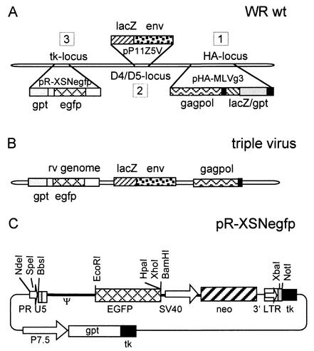
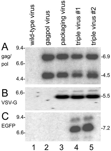
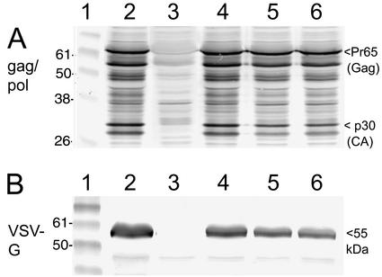
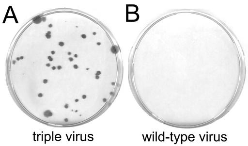
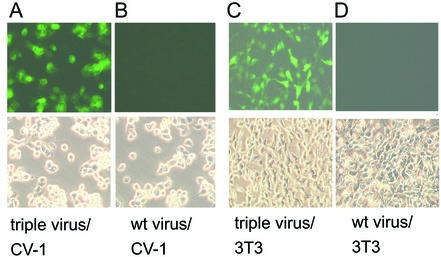
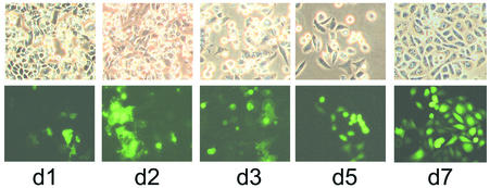
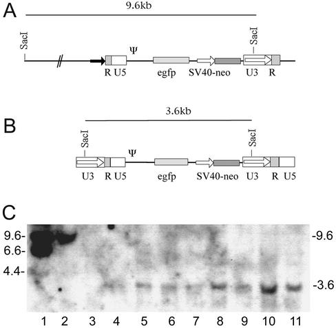
Recombinant vaccinia viruses that express defective retroviral vectors upon a single infection event in normal host cells were constructed. The gag-pol and envelope genes and a retroviral vector unit were inserted as vaccinia virus promoter-controlled transcription units at three separate loci. The triple recombinant virus was used to infect such diverse cell types as monkey and rabbit kidney, human lung, and primary chicken cells, resulting in the production of transduction-competent defective retroviral vectors. Infection of Chinese hamster ovary cells, which are nonpermissive for vaccinia virus replication, also resulted in production of retroviral vectors and secondary permanent transduction of the host cells. Since vaccinia virus supports the expression of cytotoxic proteins, the vesicular stomatitis virus G glycoprotein could be chosen as the envelope allowing a broad host range of transduction. Functionality of particles was monitored by expression of the green fluorescent protein in transduced 3T3 cell clones. This is the first description of a single chimeric virus encoding and releasing functional retroviral vectors, providing proof of principle of the new concept. No replication-competent retrovirus was detectable by sensitive reverse transcriptase assays. Since vaccinia virus has a broad host range, is extremely robust, and can be obtained at high titers and safe nonreplicating vaccinia virus strains are available, the hybrid system may open new perspectives for gene delivery.


Figures

FIG. 1.
FIG. 1.
Schematic representation of the construction of the chimeric vaccinia viruses and structure of the plasmid pR-XSNegfp. (A) In step 1, the retroviral gag-pol sequences were inserted into the vaccinia virus hemagglutinin locus (HA locus). The lacZ and gpt markers were used transiently and are not present in the first construct, the virus vHA-MLVg. The second step was insertion of the VSV-G pseudo-env gene into the D4/D5 intergenic region (D4/D5 locus), resulting in the packaging virus. The last step consisted of insertion of a retroviral vector unit into the packaging virus, resulting in the triple virus. (B) Structure of the triple virus. (C) Structure of plasmid pR-XSNegfp. This plasmid transfers the retroviral vector genome together with a permanent gpt marker into the vaccinia virus thymidine kinase (tk) locus; PR, synthetic vaccinia virus early promoter R; U5, U5 region of the retroviral long terminal repeat; Ψ, packaging signal; EGFP, open reading frame of the EGFP gene; SV40, simian virus 40 early promoter; neo, neomycin resistance gene; 3′LTR, 3′ retroviral long terminal repeat; P7.5, vaccinia virus early/late promoter P7.5; gpt, selection marker gene encoding the Escherichia coli enzyme guanine phosphoribosyltransferase.
FIG. 2.
FIG. 2.
Characterization of recombinant vaccinia viruses by Southern blotting. Genomic DNA of the different viruses was digested with the respective restriction endonuclease, transferred to a filter, and hybridized to the indicated radioactively labeled probes. (A) Viral DNAs hybridized to the gag-pol probe showed the expected MunI fragments of 6.9 and 4.5 kb in the gag-pol, packaging, and triple viruses (lanes 2 to 5). (B) Hybridization with the VSV-G probe revealed the characteristic 5.5-kb Asp718 fragments in the packaging and triple viruses (lanes 3 to 5). (C) The 7.2-kb XhoI fragment hybridizing with the EGFP probe was found only in the triple viruses (lanes 4 and 5). The wild-type virus (lanes 1) did not show these signals. The numbers at the left show the marker bands; those at the right show the sizes of the fragments (in kilobase pairs).
FIG. 3.
FIG. 3.
Expression of retroviral packaging components by recombinant vaccinia viruses shown by Western blotting. CV-1 cells were infected with the indicated viruses, and total cellular extracts were prepared and subjected to the Western blot procedure. (A) Incubation of the blots with a gag-pol antiserum revealed a characteristic pattern of protein bands, including the Pr65 gag-specific band and the p30 capsid (CA) band (arrowhead, right side) in the packaging virus and two clones of the triple viruses. Lane 1, protein markers. Lane 2, positive control, lysate of cells infected with the virus vHA-MLVg, which has a single insert of the gag-pol sequences (see text). Lane 3, negative control, lysate of cells infected with WR wild-type virus. Lane 4, lysate of cells infected with the packaging virus. Lane 5, lysate of cells infected with triple virus clone 1. Lane 6, lysate of cells infected with triple virus clone 2. (B) Incubation of blots with VSV-G antibodies revealed the typical 55-kDa band of the VSV-G glycoprotein in the packaging and triple viruses. Lane 1, protein markers. Lane 2, positive control, lysate of cells infected with the virus vDD4-mH5-VSVg, a recombinant WR-based virus with a single VSV-G gene insert. Lane 3, negative control, lysate of cells infected with wild-type WR virus. Lane 4, lysate of cells infected with the packaging virus. Lane 5, lysate of cells infected with triple virus clone 1. Lane 6, lysate of cells infected with triple virus clone 2. Numbers at the left indicate the size of the protein markers, and those at the right show the size of characteristic bands (in kilodaltons).
FIG. 4.
FIG. 4.
Colony-forming assays in NIH 3T3 cells. The kidney cell line CV-1 was infected with the indicated viruses, and the vaccinia virus-free supernatants were used to perform colony-forming assays in NIH 3T3 cells with the transduced neomycin resistance marker. Colonies were selected at 500 μg of the antibiotic G418 per ml and stained with crystal violet after 2 weeks. (A) Supernatants of CV-1 cells (the 1:1,000 dilution is shown) infected with the vaccinia virus triple virus gave rise to G418-resistant NIH 3T3 colonies. (B) Supernatants of the same cell line infected with wild-type virus did not result in colonies of the indicator cell line.
FIG. 5.
FIG. 5.
Induction of green fluorescence by infections with triple virus or transduction with retroviral particles induced by triple virus. CV-1 cells directly infected (1 PFU per cell) with the triple virus showed a strong fluorescence 12 h after infection (A), while the wild-type (wt) virus did not induce a signal (B). The characteristic cytopathic effects could be observed in the phase contrast picture (lower panels). Supernatants of CV-1 cells infected with the triple virus were used to transduce NIH 3T3 cells. Green fluorescence of NIH 3T3 clones after 6 days is shown (C). A parallel experiment with the same cells transduced with control supernatants (wild-type virus-infected CV-1 cells) did not result in a signal (D).
FIG. 6.
FIG. 6.
Infection of nonpermissive Chinese hamster ovary cells with triple virus results in secondary transductions. CHO cells were infected with 0.5 PFU of the triple virus, and green fluorescence of the culture was monitored over 7 days (d1 to d7). Phase contrast pictures (upper panel) and EGFP fluorescence (lower panel) are shown. In the first days after infection with the vaccinia virus hybrid virus (d1 to d3), infected CHO cells showed a strong cytopathic effect and also showed green fluorescence. The vaccinia virus-infected cells underwent apoptosis and detached, and after 5 days (d5), the noninfected but transduced cells recovered and formed confluent monolayers with islands of fluorescing cells (d7).
FIG. 7.
FIG. 7.
(A) Structure of the retroviral vector unit integrated in the triple virus and in virus vrR-XSNegfp. Since the 5′ LTR was modified (the U3 region was replaced with a vaccinia virus early promoter; black arrow), the SacI site usually present in the 5′ LTR was deleted. A SacI site is located in the vaccinia virus genome approximately 6 kb upstream of the integration site of the retroviral vector unit, resulting in the 9.6-kb viral SacI fragment. (B) Structure of the provirus in the transduced cell lines. During transduction, the 5′ LTR is reconstituted in the reverse transcription process, and the SacI site reappears, resulting in the 3.6-kb SacI fragment found in the transduced cell lines. (C) Genomic Southern blots of G418-positive NIH 3T3 clones transduced with cell culture supernatants obtained after infection of different cell types. Clones obtained after transduction with retroviral particles produced in CHO cells (lanes 4 and 5), CV-1 cells (lanes 6 and 7), Vero cells (lanes 8 and 9), and chicken cells (lanes 10 and 11) showed the characteristic 3.6-kb SacI band that indicates an integrated retroviral vector. Lane 1, marker DNA; lane 2, DNA of the virus vrR-XSNegfp harboring only the retroviral vector unit; lane 3, DNA of a nontransduced NIH 3T3 clone. Numbers at the left show the size of the marker bands; numbers at the right show size of the hybridizing bands in the samples (in kilobase pairs).

Similar articles

Cited by

References

    1. Burns, J. C., T. Friedmann, W. Driever, M. Burrascano, and J. K. Yee. 1993. Vesicular stomatitis virus G glycoprotein pseudotyped retroviral vectors: concentration to very high titer and efficient gene transfer into mammalian and nonmammalian cells. Proc. Natl. Acad. Sci. USA 90:8033-8037. - PMC - PubMed
    1. Chakrabarti, S., K. Brechling, and B. Moss. 1985. Vaccinia virus expression vector: coexpression of β-galactosidase provides visual screening of recombinant virus plaques. Mol. Cell. Biol. 5:3403-3409. - PMC - PubMed
    1. Chakrabarti, S., J. R. Sisler, and B. Moss. 1997. Compact, synthetic, vaccinia virus early/late promoter for protein expression. BioTechniques 23:1094-1097. - PubMed
    1. Falkner, F. G., and B. Moss. 1988. Escherichia coli gpt gene provides dominant selection for vaccinia virus open reading frame expression vectors. J. Virol. 62:1849-1854. - PMC - PubMed
    1. Feng, M., W. H. Jackson, Jr., C. K. Goldman, C. Rancourt, M. Wang, S. K. Dusing, G. Siegal, and D. T. Curiel. 1997. Stable in vivo gene transduction via a novel adenoviral/retroviral chimeric vector. Nat. Biotechnol. 15:866-870. - PubMed

Publication types

Substances

LinkOut - more resources