. 2003 Jun;77(12):7034-40.
doi: 10.1128/jvi.77.12.7034-7040.2003.

Intraventricular brain injection of adeno-associated virus type 1 (AAV1) in neonatal mice results in complementary patterns of neuronal transduction to AAV2 and total long-term correction of storage lesions in the brains of beta-glucuronidase-deficient mice

Affiliations

Intraventricular brain injection of adeno-associated virus type 1 (AAV1) in neonatal mice results in complementary patterns of neuronal transduction to AAV2 and total long-term correction of storage lesions in the brains of beta-glucuronidase-deficient mice

Marco A Passini et al. J Virol. 2003 Jun.

Abstract

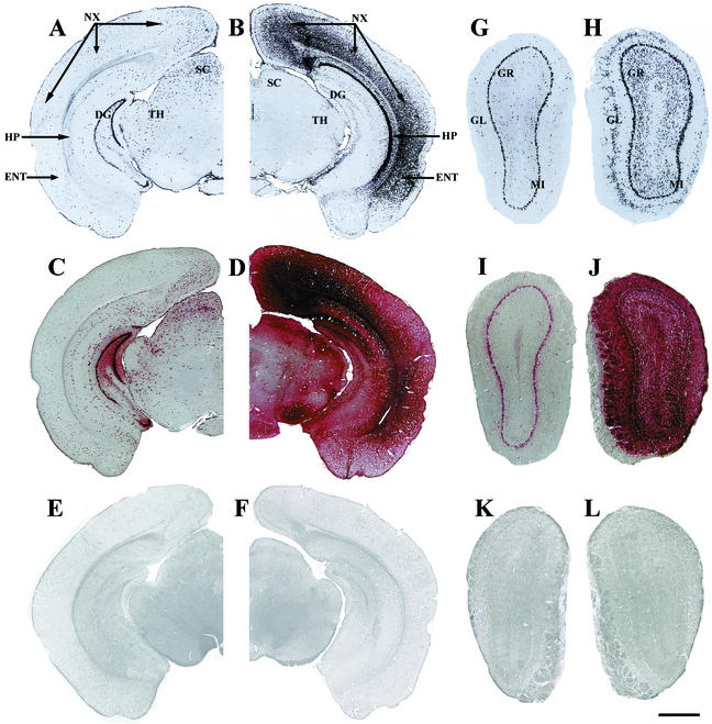
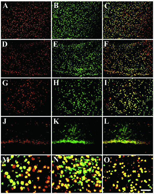
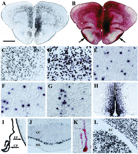
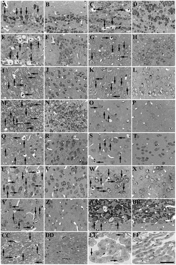
Inherited metabolic disorders that affect the central nervous system typically result in pathology throughout the brain; thus, gene therapy strategies need to achieve widespread delivery. We previously found that although intraventricular injection of the neonatal mouse brain with adeno-associated virus serotype 2 (AAV2) results in dispersed gene delivery, many brain structures were poorly transduced. This limitation may be overcome by using different AAV serotypes because the capsid proteins use different cellular receptors for entry, which may allow enhanced global targeting of the brain. We tested this with AAV1 and AAV5 vectors. AAV5 showed very limited brain transduction after neonatal injection, even though it has different transduction patterns than AAV2 in adult brain injections. In contrast, AAV1 vectors, which have not been tested in the brain, showed robust widespread transduction. Complementary patterns of transduction between AAV1 and AAV2 were established and maintained in the adult brain after neonatal injection. In the majority of structures, AAV1 transduced many more cells than AAV2. Both vectors transduced mostly neurons, indicating that differential expression of receptors on the surfaces of neurons occurs in the developing brain. The number of cells positive for a vector-encoded secreted enzyme (beta-glucuronidase) was notably greater and more widespread in AAV1-injected brains. A comprehensive analysis of AAV1-treated brains from beta-glucuronidase-deficient mice (mucopolysaccharidosis type VII) showed complete reversal of pathology in all areas of the brain for at least 1 year, demonstrating that the combination of this serotype and experimental strategy is therapeutically effective for treating global neurometabolic disorders.


Figures

FIG. 1.
FIG. 1.
Complementary patterns of transduction after intraventricular injection of normal neonatal mice with AAV1-HβH or AAV2-HβH at 1 month p.i. Expression of virally encoded mRNA was detected by in situ hybridization with an antisense riboprobe against the human GUSB cDNA (A, B, G, and H), and biologically active enzyme was detected by histochemistry (C, D, I, and J). The AAV2 transduction patterns shown here (A, C, G, and I) were previously published (25) and are presented as a direct comparison to AAV1. (A) A section through the caudal forebrain-rostral midbrain showed AAV2 transduction in the neocortex, entorhinal cortex, superior colliculus, and dentate gyrus and in the thalamus and pretectal nucleus. (B) In comparison, AAV1 produced substantially higher numbers of in situ hybridization-positive cells in the CA1 to CA3 pyramidal and oriens cell layers of the hippocampus and in the neocortex and entorhinal cortex but produced smaller numbers of in situ hybridization-positive cells in the dentate gyrus, superior colliculus, and thalamus and pretectal nucleus. (C) Enzyme-positive cells were relatively confined to transduced cells in AAV2-injected brains. (D) In contrast, there was enzyme spread throughout the entire brain with AAV1. (E and F) The uninjected, normal control brain was not positive for GUSB activity in the absence (E) or presence (F) of heat inactivation. (G to J) Complementary patterns of transduction also occurred in other regions of the brain, such as the olfactory bulb. (G and I) With AAV2-HβH, transduction and enzymatic activity were detected mostly in the mitral cell layer. (H) In contrast, AAV1 produced robust transduction in the mitral, glomerular, and granule cell layers throughout the entire olfactory bulb. (J) This increase in transduction resulted in large amounts of biologically active enzyme in all laminar layers of the olfactory bulb. (K and L) The uninjected, normal control olfactory bulb did not show GUSB activity in the absence (K) or presence (L) of heat inactivation. Abbreviations: DG, dentate gyrus; ENT, entorhinal cortex; GL, glomerular cell layer; GR, granule cell layer; HP, CA1 to CA3 areas of the hippocampus; MI, mitral cell layer; NX, neocortex; SC, superior colliculus; TH, thalamus and pretectal nucleus. Bar: 1,000 μm (A to D) or 500 μm (E to H).
FIG. 2.
FIG. 2.
AAV1-HβH is a neurotropic vector. Fluorescent in situ hybridization with the human GUSB riboprobe (red signal) (A, D, G, and J), immunofluorescence against neuron-specific enolase (green signal) (B, E, H, and K), and program overlay (yellow signal) (C, F, I, and L to O). The neocortex (A to C and M), entorhinal cortex (D to F and N), striatum (G to I and O), and CA1 cell layer of the hippocampus (J to L) of normal mice at 1 month p.i. Bar: 200 μm (A to L) or 50 μm (M to O).
FIG. 3.
FIG. 3.
Long-term, widespread transduction with AAV1-HβH in the brains of normal mice at 1 year p.i. as determined by in situ hybridization (A, C to J, and L) and enzyme histochemistry (B and K). (A) Transduction was observed throughout the rostral forebrain. (B) Enzyme-positive cells were detected in transduced sites and in regions not positive for GUSB mRNA, such as the myelinated lateral olfactory tracts (arrows). Other regions that maintained expression included the striatum (C), entorhinal cortex (D), inferior colliculus (E), amygdala (F), hypothalamus (G), third ventricle and the surrounding periventricular region (H), ependyma and choroid plexus (I), and the Purkinje cell layer of the cerebellum (J). (K) Histochemistry showed an enzyme-positive cell with Purkinje cell morphology. (L) None of the white-matter tracts were transduced, such as the external capsule. Abbreviations: CP, choroid plexus; EC, external capsule; EP, ependyma; GC, granule cell layer of the cerebellum; ML, molecular layer of the cerebellum; PJ, Purkinje cell layer. Bar: 1,000 μm (A and B), 500 μm (H), 250 μm (C, I, J, and L), 125 μm (D), or 60 μm (E to G and K).
FIG.4.
FIG.4.
Reversal of storage lesions in MPS VII brains 1 year after intraventricular injection of AAV1-HβH. Uninjected MPS VII control (A, C, E, G, I, K, M, O, Q, S, U, W, Y, AA, CC, and EE) and AAV1 vector-injected MPS VII (B, D, F, H, J, L, N, P, R, T, V, X, Z, BB, DD, and FF) brains. The mitral (A and B), granule (C and D), and glomerular (E and F) cell layers of the olfactory bulb; the piriform cortex (G and H); the neocortex (I and J); the entorhinal cortex (K and L); the CA3 pyramidal cell layer of the hippocampus (M and N); the striatum (O and P); the hypothalamus (Q and R); the amygdala (S and T); the subiculum (U and V); the thalamus (W and X); the inferior colliculus (Y and Z); the Purkinje cell layer in the cerebellum (AA and BB); the external capsule (CC and DD); and the choroid plexus (EE and FF) are shown. Arrows point to representative neurons, glia, and microglia with lysosomal storage vacuoles. Bar: 25 μm.

Similar articles

Cited by

References

    1. Alisky, J. M., S. M. Hughes, S. L. Sauter, D. Jolly, T. W. Dubensky, P. D. Staber, J. A. Chiorini, and B. L. Davidson. 2000. Transduction of murine cerebellar neurons with recombinant FIV and AAV5 vectors. Neuroreport 1:2669-2673. - PubMed
    1. Auricchio, A., G. Kobinger, V. Anand, M. Hildinger, E. O'Connor, A. M. Maguire, J. M. Wilson, and J. Bennett. 2001. Exchange of surface proteins impacts on viral vector cellular specificity and transduction characteristics: the retina as a model. Hum. Mol. Genet. 10:3075-3081. - PubMed
    1. Bantel-Schaal, U., H. Delius, R. Schmidt, and H. Van Hausen. 1999. Human adeno-associated virus type 5 is only distantly related to other known primate helper-dependent parvovirus. J. Virol. 73:939-947. - PMC - PubMed
    1. Barthel, L. K., and P. A. Raymond. 2000. In situ hybridization studies of retinal neurons. Methods Enzymol. 316:579-590. - PubMed
    1. Bartlett, J. S., R. J. Samulski, and T. J. McCown. 1998. Selective and rapid uptake of adeno-associated virus type 2 in brain. Hum. Gene Ther. 9:1181-1186. - PubMed

Publication types

MeSH terms