. 2003 Jun;77(12):7041-7.
doi: 10.1128/jvi.77.12.7041-7047.2003.

Interactions of rotavirus VP4 spike protein with the endosomal protein Rab5 and the prenylated Rab acceptor PRA1

Affiliations

Interactions of rotavirus VP4 spike protein with the endosomal protein Rab5 and the prenylated Rab acceptor PRA1

Vincent Enouf et al. J Virol. 2003 Jun.

Abstract

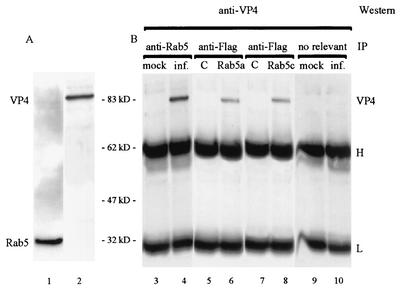
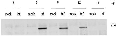
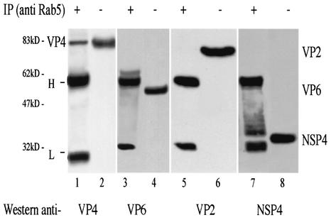
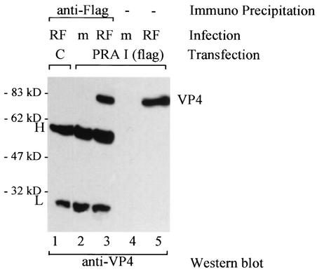
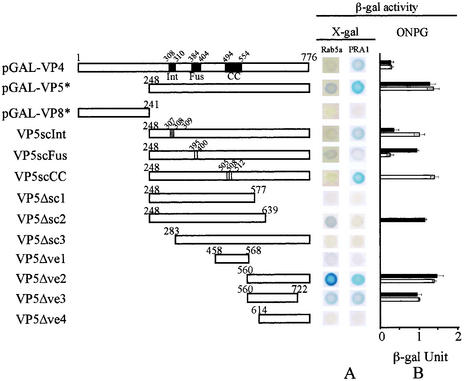
Rotavirus spike protein VP4 is implicated in several important functions, such as cell attachment, penetration, hemagglutination, neutralization, virulence, and host range. It is present at the plasma membrane and colocalizes with the cytoskeleton in infected cells. We looked for cellular partners responsible for the localization of VP4 by two-hybrid screening of a monkey CV1 cell cDNA library. In the screen we isolated repeatedly three cDNAs encoding either two isoforms (a and c) of Rab5 protein or the prenylated Rab acceptor (PRA1). The small GTPase Rab5 is a molecule regulating the vesicular traffic and the motility of early endosomes along microtubules. Rab5 interacts with a large number of effectors, in particular with PRA1. Interactions of VP4 with both partners, Rab5 and PRA1, were confirmed by coimmunoprecipitation from infected- or transfected-cell lysates. Interaction of Rab5 and PRA1 was restricted to free VP4, since neither triple-layered particles nor NSP4-VP4-VP7 heterotrimeric complexes could be coprecipitated. Site-directed and deletion mutants of VP4 were used to map a VP4 domain(s) interacting with Rab5 or PRA1. Of the 10 mutants tested, 2 interacted exclusively with a single partner. In contrast, the domain extending from amino acids 560 to 722 of VP4 is essential for both interactions. These results suggest that Rab5 and PRA1 may be involved in the localization and trafficking of VP4 in infected cells.


Figures

FIG. 1.
FIG. 1.
Coimmunoprecipitation of VP4 and Rab5 protein in infected MA104 and transfected COS-7 cells. (A) Lysate from infected MA104 cells at 6 h p.i. was analyzed by SDS-PAGE and anti-Rab5 or anti-VP4 Western blotting (lanes 1 and 2, respectively). (B) Lysates from infected (inf.) or mock-infected (mock) MA104 cells were subjected to immunoprecipitation with protein A-Sepharose beads loaded with anti-Rab5 MAb (lanes 3 and 4) or nonrelevant antibody (lanes 9 and 10). Immunoprecipitates were analyzed by SDS-PAGE and anti-VP4 Western blotting. Lysates for infected and transfected COS-7 cells with pcDNA3.1-Rab5a (Rab5a), pcDNA3.1-Rab5c (Rab5c), and pcDNA3.1 (C) were subjected to immunoprecipitation with protein A-Sepharose beads loaded with anti-FLAG MAb. Immunoprecipitates were analyzed by SDS-PAGE and anti-VP4 Western blotting. Molecular mass markers are indicated between panels A and B. H and L correspond to heavy and light chains of immunoglobulin G, respectively.
FIG. 2.
FIG. 2.
Time course of interaction between VP4 and Rab5. Lysates were prepared from mock-infected (mock) or infected (inf.) MA104 cells at 3, 6, 9, 12, and 18 h after rotavirus infection. Each lane corresponds to the total protein content of 9 × 10⁶ cells. These lysates were subjected to immunoprecipitation with protein A-Sepharose beads loaded with anti-Rab5 MAb. Immunoprecipitates were analyzed by SDS-PAGE and anti-VP4 Western blotting.
FIG. 3.
FIG. 3.
Interaction among rotavirus proteins VP2, VP6, NSP4, and Rab5. Lysate of infected MA104 cells 6 h p.i. was subjected to immunoprecipitation with protein A-Sepharose beads loaded with anti-Rab5 MAb. Immunoprecipitates were analyzed by SDS-PAGE and anti-VP4, anti-VP6, anti-VP2, or anti-NSP4 Western blotting (lanes 1, 3, 5, and 7, respectively). The same lysates were analyzed directly, i.e., prior to immunoprecipitation, by SDS-PAGE and anti-VP4, anti-VP6, anti-VP2, or anti-NSP4 Western blotting (lanes 2, 4, 6, and 8, respectively). Each lane corresponds to the total protein content of 9 × 10⁶ cells. H and L correspond to heavy and light chains of immunoglobulin G, respectively. The additional band present in lane 7 is probably due to a contaminant protein in the anti-Rab5 antibodies. Migration of molecular mass markers is indicated on the left.
FIG. 4.
FIG. 4.
Interaction between VP4 and PRA1. Lysates from infected (RF) or mock-infected (m) COS-7 cells transfected with pcDNA3.1-PRA1 were subjected to immunoprecipitation with protein A-Sepharose beads loaded with antibodies specific for FLAG. A lysate of infected and transfected COS-7 cells with pcDNA3.1 Hygro-FLAG vector was used as a control and submitted to the same immunoprecipitation procedure (lane 1). Immunoprecipitates were analyzed by SDS-PAGE and anti-VP4 Western blotting. Total lysates from infected (RF) or from mock-infected (m) COS-7 cells transfected with pcDNA3.1-PRA1 were analyzed by SDS-PAGE and anti-VP4 Western blotting (lanes 4 and 5). Each lane corresponds to the total protein content of 9 × 10⁶ cells. H and L correspond to heavy and light chains of immunoglobulin G, respectively. Migration of molecular mass markers is indicated on the left.
FIG. 5.
FIG. 5.
Schematic diagram of the truncated VP4 and VP5* proteins fused to the GAL4 binding domain and analysis of VP4-Rab5 and VP4-PRA1 interactions. (A) Bars represent the protein product of each deleted VP4 gene, with amino acid positions indicated. Positions of amino acid changes are indicated for VP5scFus, VP5scInt, and VP5scCC mutants. Int, integrin receptor; Fus, fusogen domain; CC, coiled-coil sequence. HF7C yeast was cotransformed with the indicated mutant and pGAD-Rab5a and pGAD-PRA1. The interaction was assessed by checking for blue colonies in the presence of X-Gal. (B) Quantitative evaluations of the interactions were obtained from independent yeast cotransformants assayed with ONPG as substrate. β-Galactosidase activity was expressed in units and calculated by the following formula: (1,000 A420)/(A600TV), where A420 is the absorbance of the reaction mixture, A600 is the cell density of the culture, T is the reaction time (in minutes), and V is the volume (in milliliters) used for the assay. Solid and open bars correspond to interactions of Rab5 and PRA1 with constructs indicated, respectively. Standard deviations are shown (n = 3).

Similar articles

Cited by

References

    1. Abdul-Ghani, M., P. Y. Gougeon, D. C. Prosser, L. F. Da-Silva, and J. K. Ngsee. 2001. PRA isoforms are targeted to distinct membrane compartments. J. Biol. Chem. 276:6225-6233. - PubMed
    1. Alory, C., and W. E. Balch. 2000. Molecular basis for Rab prenylation. J. Cell Biol. 150:89-103. - PMC - PubMed
    1. Altschul, S. F., T. L. Madden, A. A. Schaffer, J. Zhang, Z. Zhang, W. Miller, and D. J. Lipman. 1997. Gapped BLAST and PSI-BLAST: a new generation of protein database search programs. Nucleic Acids Res. 25:3389-3402. - PMC - PubMed
    1. Belin, M. T., and P. Boulanger. 1993. Involvement of cellular adhesion sequences in the attachment of adenovirus to the HeLa cell surface. J. Gen. Virol. 74:1485-1497. - PubMed
    1. Bennett, M. K., and R. H. Scheller. 1993. The molecular machinery for secretion is conserved from yeast to neurons. Proc. Natl. Acad. Sci. USA 90:2559-2563. - PMC - PubMed

LinkOut - more resources