. 2003 Jun;77(12):7078-92.
doi: 10.1128/jvi.77.12.7078-7092.2003.

Basic residues of the helix six domain of influenza virus M1 involved in nuclear translocation of M1 can be replaced by PTAP and YPDL late assembly domain motifs

Affiliations

Basic residues of the helix six domain of influenza virus M1 involved in nuclear translocation of M1 can be replaced by PTAP and YPDL late assembly domain motifs

Eric Ka-Wai Hui et al. J Virol. 2003 Jun.

Retraction in

  • Retraction.
    Hui EK, Barman S, Yang TY, Tang DH, France B, Nayak DP. Hui EK, et al. J Virol. 2006 Oct;80(20):10289. doi: 10.1128/JVI.01632-06. J Virol. 2006. PMID: 17005709 Free PMC article. No abstract available.

Abstract

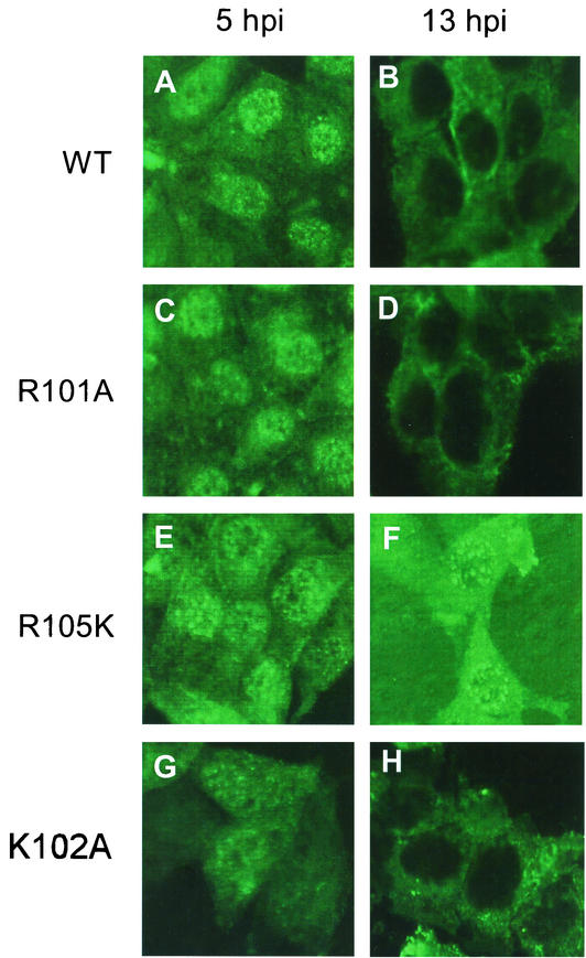
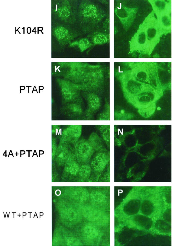
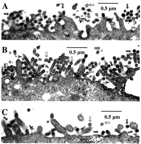
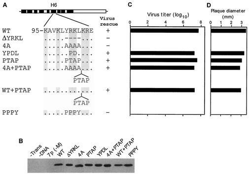
Influenza type A virus matrix (M1) protein possesses multiple functional motifs in the helix 6 (H6) domain (amino acids 91 to 105), including nuclear localization signal (NLS) (101-RKLKR-105) involved in translocating M1 from the cytoplasm into the nucleus. To determine the role of the NLS motif in the influenza virus life cycle, we mutated these and the neighboring sequences by site-directed mutagenesis, and influenza virus mutants were generated by reverse genetics. Our results show that infectious viruses were rescued by reverse genetics from all single alanine mutations of amino acids in the H6 domain and the neighboring region except in three positions (K104A and R105A within the NLS motif and E106A in loop 6 outside the NLS motif). Among the rescued mutant viruses, R101A and R105K exhibited reduced growth and small-plaque morphology, and all other mutant viruses showed the wild-type phenotype. On the other hand, three single mutations (K104A, K105A, and E106A) and three double mutations (R101A/K102A, K104A/K105A, and K102A/R105A) failed to generate infectious virus. Deletion (Delta YRKL) or mutation (4A) of YRKL also abolished generation of infectious virus. However, replacement of the YRKL motif with PTAP or YPDL as well as insertion of PTAP after 4A mutation yielded infectious viruses with the wild-type phenotype. Furthermore, mutant M1 proteins (R101A/K102A, Delta YRKL, 4A, PTAP, 4A+PTAP, and YPDL) when expressed alone from cloned cDNAs were only cytoplasmic, whereas the wild-type M1 expressed alone was both nuclear and cytoplasmic as expected. These results show that the nuclear translocation function provided by the positively charged residues within the NLS motif does not play a critical role in influenza virus replication. Furthermore, these sequences of H6 domain can be replaced by late (L) domain motifs and therefore may provide a function similar to that of the L domains of other negative-strand RNA and retroviruses.


Figures

FIG. 1.
FIG. 1.
(A) Schematic diagram of influenza A virus M1 with functional domains. α-Helix 1 (H1) to helix 9 (H9) (black boxes) and intervening loops (L) are shown according to the work of Sha and Luo (59). The hydrophobic regions between aa 1 and 11, 62 and 68, and 114 and 133 are shown as lipid binding domains (LBD) (17, 18, 71), between aa 91 and 111 as RNA binding domain (65, 67), aa 90 and 110 and aa 129 and 164 as TID (45, 65, 69, 71), aa 101 and 105 as NLS (69, 72), and aa 148 and 162 as predicted CCHH zinc finger motif (Zn) (64). The interaction of M1 with NS2 or RNP is located within the C-terminal two-thirds (68, 71). The potential protein kinase C phosphorylation sites (circled P) are Ser70, Ser161, and Thr185 (51). (B) Amino acid sequence of the H6 domain (aa 91 to 105) and 106E. The positively charged amino acids are boxed in gray. Schematic diagram of the helix (H1 to H9) and loops. H6 is exposed on the surface (3, 20, 21, 59). The α-helical regions are indicated as cylinders, and the putative N and C termini are marked. (C) Helical wheel plot of H6 domain. Positively charged amino acids (gray shading) are exposed on the surface. (D) Amino acid sequence alignment of the H6 domains of M1 from different influenza A virus strains. The positively charged amino acids are boxed in gray. Variations are shown.
FIG. 2.
FIG. 2.
(A) Schematic representation of single-amino-acid mutations (aa 95 to 106). Amino acid sequence of the H6 domain (aa 95 to 105) and specific mutations are indicated. The positively charged amino acids are shown in gray. (B) Immunoprecipitation of M1 mutant proteins. At 18 h posttransfection, 293T cells were pulse labeled for 2 h. Cells were then lysed in RIPA buffer, immunoprecipitated with anti-M1 antibody, and resolved by SDS-12% PAGE. Autoradiographs from two separate gels are shown. (C) PFU titer of transfectant viruses. Transfected viruses were rescued by eight plasmids in transfection of 293T cells. MDCK cells were then infected with transfectant virus at a MOI of 0.001 and maintained in VGM containing 0.5 μg of trypsin/ml as described in Materials and Methods. Supernatants were collected at 48 h p.i. and assayed for numbers of PFU by plaque assay. Data represent mean values (n = 3). Asterisk, P < 0.001 (versus WT). (D) The plaque sizes for different mutant viruses on MDCK cells. Plaques were visualized at day 3, and diameters were measured. Data represent mean values (n = 4). Asterisk, P < 0.001 (versus WT).
FIG. 3.
FIG. 3.
(A) Schematic representation of the double-mutation constructs. (B) Immunoprecipitation and PAGE analysis of M1 mutant proteins. (C) Titer of transfectant viruses. (D) Plaque sizes for different mutants on MDCK cells. For details, see the legend to Fig. 2.
FIG. 4.
FIG. 4.
Stability of WT and R101A and R105K mutated M1 proteins. At 18 h posttransfection, 293T cells were pulse labeled for 2 h (lanes P) and then chased with excess Met and Cys for 3 h (lanes C). Cells were then lysed in RIPA buffer, immunoprecipitated with anti-M1 antibody, and analyzed by SDS-PAGE.
FIG. 5.
FIG. 5.
Intracellular distribution of M1 mutants by IF analysis of virus-infected MDCK cells. MDCK cells were grown on slides and infected with WT or mutant viruses at a MOI of 3.0. At 5 h p.i. (panels A, C, E, G, I, K, M, and O) or 13 h p.i. (panels B, D, F, H, J, L, N, and P), the cells were fixed and stained for M1 by IF with an anti-M1 antibody. (A and B) WT; (C and D) R101A; (E and F) R105K; (G and H) K102A; (I and J) K104R; (K and L) PTAP; (M and N) 4A+PTAP; (O and P) WT+PTAP. Representative fields from analysis of 20 fields of view in duplicate experiments are shown. Magnification, ×558 (A-I) and 527 (I-P).
FIG. 5.
FIG. 5.
Intracellular distribution of M1 mutants by IF analysis of virus-infected MDCK cells. MDCK cells were grown on slides and infected with WT or mutant viruses at a MOI of 3.0. At 5 h p.i. (panels A, C, E, G, I, K, M, and O) or 13 h p.i. (panels B, D, F, H, J, L, N, and P), the cells were fixed and stained for M1 by IF with an anti-M1 antibody. (A and B) WT; (C and D) R101A; (E and F) R105K; (G and H) K102A; (I and J) K104R; (K and L) PTAP; (M and N) 4A+PTAP; (O and P) WT+PTAP. Representative fields from analysis of 20 fields of view in duplicate experiments are shown. Magnification, ×558 (A-I) and 527 (I-P).
FIG. 6.
FIG. 6.
Budding of virus particles by thin-section electron microscopy. MDCK cells grown on polycarbonate filter were infected with either WT, K101A, or K105R viruses at a MOI of 3.0. At 12 h p.i., infected cell monolayers on filters were cross-linked, postfixed, and embedded. Sixty-nanometer-thick sections were stained and examined. WT (A), R101A (B), and R105K (C). ➞, normal virus particles; ➱, empty VLPs; →, elongated virus particles.
FIG. 7.
FIG. 7.
Protein composition of purified WT, R101A, or R105K virion. Virus-infected MDCK cells were labeled from 4 to 16 h p.i. with 250 μCi of 35S-protein label. Supernatants were harvested and clarified, and labeled viruses in supernatants were pelleted through a 25% sucrose cushion by centrifugation. The pelleted particles were resuspended with TNE buffer, lysed in RIPA buffer with 1% SDS at 37°C for 90 min, and directly analyzed by SDS-12% PAGE. The position of viral proteins HA, NP, and M1 are indicated at the left of panel. The ratio based on HA, NP, or M1 is mentioned at the bottom of the gel. The data represent the average for three independent experiments with less than 10% variation.
FIG. 8.
FIG. 8.
(A) Schematic representation of the YRKL mutation, deletion, replacement, or insertion constructs. Different L domain motifs were used for replacement or insertion. (B) Immunoprecipitation of M1 mutant proteins. (C) Virus titer of transfectant viruses. (D) Plaque sizes for different mutants on MDCK cells. For details, see the legend to Fig. 2.
FIG.9.
FIG.9.
Intracellular distribution of M1 mutants by IF in cDNA-transfected 293T cells. 293T cells were transfected with WT or mutant M1 cDNA. At 8 h posttransfection, cells were fixed and stained for M1 by IF with an anti-M1 antibody. (A) WT; (B) K104A; (C) R105A; (D) Y100A/L103A; (E) R101A/K102A; (F) 4A; (G) ΔYRKL; (H) PTAP; (I) 4A+PTAP; (J) YPDL; (K) PPPY. Representative fields from analysis of 20 fields of view in duplicate experiments are shown. Magnification, ×558.

Similar articles

Cited by

References

    1. Accola, M. A., B. Strack, and H. G. Göttlinger. 2000. Efficient particle production by minimal Gag constructs which retain the carboxy-terminal domain of human immunodeficiency virus type 1 capsid-p2 and a late assembly domain. J. Virol. 74:5395-5402. - PMC - PubMed
    1. Allen, H., J. McCauley, M. Waterfield, and M. J. Gething. 1980. Influenza virus RNA segment 7 has the coding capacity for two polypeptides. Virology 107:548-551. - PubMed
    1. Arzt, S., F. Baudin, A. Barge, P. Timmins, W. P. Burmeister, and R. W. H. Ruigrok. 2001. Combined results from solution studies on intact influenza virus M1 protein and from a new crystal form of its N-terminal domain show that M1 is an elongated monomer. Virology 279:439-446. - PubMed
    1. Barman, S., L. Adhikary, Y. Kawaoka, and D. P. Nayak. 2003. Influenza A virus hemagglutinin containing basolateral localization signal does not alter the apical budding of a recombinant influenza A virus in polarized MDCK cells. Virology 305:138-152. - PubMed
    1. Baudin, F., C. Bacj, S. Cusak, and R. W. H. Ruigrok. 1994. Structure of influenza virus RNP. I. Influenza virus nucleoprotein melts secondary structure in panhandle RNA and exposes the bases to the solvent. EMBO J. 13:3158-3165. - PMC - PubMed

Publication types

MeSH terms

Substances

LinkOut - more resources