. 2003 Jun;77(12):7106-12.
doi: 10.1128/jvi.77.12.7106-7112.2003.

Activated Notch1 inhibits p53-induced apoptosis and sustains transformation by human papillomavirus type 16 E6 and E7 oncogenes through a PI3K-PKB/Akt-dependent pathway

Affiliations

Activated Notch1 inhibits p53-induced apoptosis and sustains transformation by human papillomavirus type 16 E6 and E7 oncogenes through a PI3K-PKB/Akt-dependent pathway

Pradip Nair et al. J Virol. 2003 Jun.

Abstract

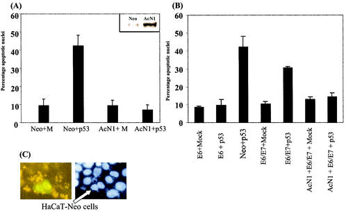
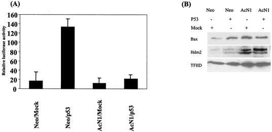
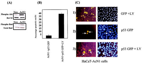
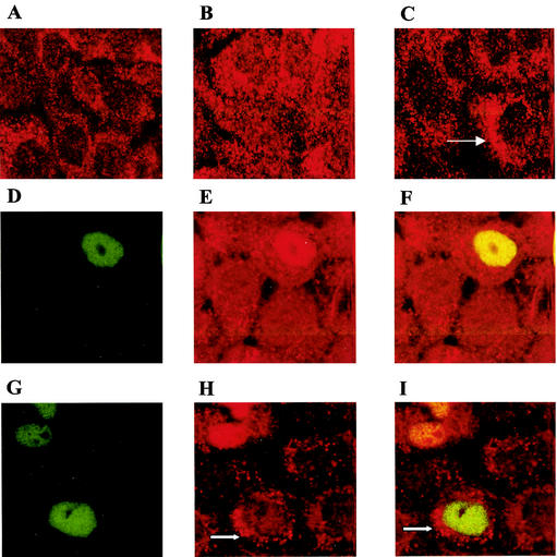
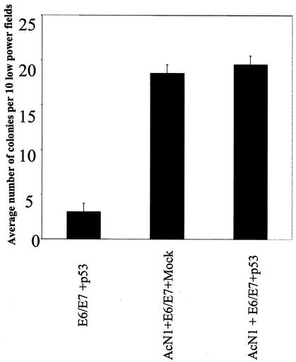
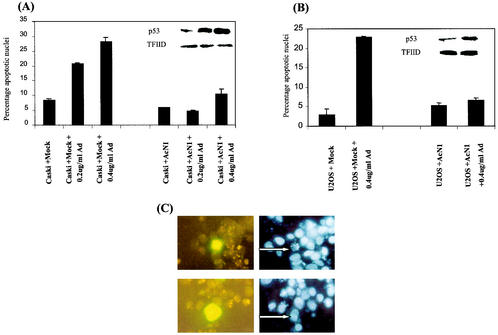
Activated Notch1 (AcN1) alleles cooperate with oncogenes from DNA tumor viruses in transformation of epithelial cells. AcN1 signaling has pleiotropic effects, and suggested oncogenic roles include driving proliferation through cyclin D1 or the generation of resistance to apoptosis on matrix withdrawal through a phosphatidylinositol 3-kinase (PI3K)-PKB/Akt-dependent pathway. Here, we extend the antiapoptotic role for AcN1 by showing inhibition of p53-induced apoptosis and transactivation. Chemical inhibitors of the PI3K pathway block AcN1-induced inhibition of p53-dependent apoptosis and nuclear localization of Hdm2. We show that expression of wild-type p53 does not inhibit synergistic transformation by AcN1 and human papillomavirus E6 and E7 oncogenes. We suggest that activation of Notch signaling may serve as an additional mechanism to inhibit wild-type p53 function in papillomavirus-associated neoplasia.


Figures

FIG. 1.
FIG. 1.
(A and B) Graphs showing percentages of apoptotic nuclei in transfected cells. (A) pCDNA3 (Mock [M]) and pCDNA3-AcN1 (AcN1) were used to generate HaCaT-Neo and HaCaT-AcN1 cells, respectively, by selecting with G-418 and pooling different clones. Inset, Notch1 overexpression in HaCaT-AcN1 stable cell lines compared to that in Neo-expressing cells. These cells were transfected with various combinations of plasmids. Mock (pCDNA3) or pCEP4-p53 along with pEGFPN1 was transfected in a ratio of 5:1 (2.5 μg of Mock or p53 plasmid with 0.5 μg of tracer GFP plasmid). GFP-positive cells were counted 24 h after transfection, counterstained with bisbenzamide (Hoechst), and assessed microscopically for nuclear condensation and chromatin fragmentation. Typically 100 GFP-positive cells were scored for apoptotic features. (B) HaCaT-E6 (lanes 1 and 2 [counting from the left]) and HaCaT-E6 and -E7 (lanes 4 and 5) cells expressing HPV-16 oncogenes were transfected with Mock (pCDNA3) or pCEP4-p53 along with pEGFPN1. HaCaT-AcN1 (lanes 6 and 7) cells were transfected with E6 and E7 plasmids along with Mock (pCDNA3) or pCEP4-p53. Standard deviations from the means of at least three independent experiments are shown for panels A and B. (C) HaCaT-Neo cells were transiently transfected with AcN1-GFP (fusion of AcN1 and GFP coding sequences in pEGFPN1 [gift from A. Sarin and H. Sade, National Centre for Biological Sciences], which was generated from a plasmid expressing AcN1 given by A. Rangarajan, Whitehead Institute) and the p53 vector in a ratio of 1:5 (0.5 μg of AcN1-GFP and 2.5 μg of p53). Adherent cells 14 h after transfection were fixed with 4% paraformaldehyde and observed under the fluorescence microscope. Ten minutes before fixation, bisbenzamide was added to the medium and the mixture was incubated at 37°C (Hoechst). Cells that were AcN1-GFP positive (nuclear GFP; arrow) were resistant to p53-induced cell death, while the controls transfected with pEGFPN1 and p53 showed around 40% cell death.
FIG. 2.
FIG. 2.
(A) Activation of the p53-dependent PG13-Luc reporter. HaCaT-Neo and HaCaT-AcN1 were transfected with either Mock (pCDNA3) or pCEP4-p53 (2 μg of DNA) along with the p53 reporter (0.5 μg of DNA). Renilla luciferase (0.2 μg of DNA) was used to normalize for transfection. Error bars, standard deviations from the means for three independent experiments. The assay was done 48 h after transfection. (B) Western blot showing Bax (N-20 [Santa Cruz Biotechnology]) and Hdm2 (AB-1 [Oncogene]) (Hdm2 dimer at 95 kDa) protein expression. Briefly, cells were lysed with 1% sodium dodecyl sulfate-1.0 mM sodium vanadate in 10 mM Tris (pH 7.4) lysis buffer heated to 95°C. Fifty micrograms of total protein, by the Bradford estimation method, was loaded into each well of a 10% polyacrylamide gel. Plasmids (1.5 μg of Mock [Neo] and p53 plasmids) were transfected in Neo- and AcN1-expressing cells. TFIID (SI-1 [Santa Cruz Biotechnology]) was used as a loading control.
FIG. 3.
FIG. 3.
Blocking PI3K eliminates resistance to p53-induced apoptosis. (A) Western blot showing endogenous phospho-Akt (antibody against Ser472, Ser473, and Ser474 [BD transLab]), phospho-Bad (antibody against Ser136 [CST]), total Bad (CST), and Bcl-Xl (S-18 [Santa Cruz Biotechnology]) protein levels in HaCaT-Neo and HaCaT-AcN1 cell lysates. (B and C) PI3K inhibitor LY294002 eliminates resistance to p53-induced apoptosis. (B) Histogram showing apoptotic nuclei from HaCaT-AcN1 cells transfected with p53-GFP and treated with LY294002. Adherent cells were transfected with p53-GFP and treated as in Fig. 1C. (C) Representative images of the histogram in panel B. Neither pEGFPN1 (section 1) nor p53-GFP (section 2) induces apoptosis (arrows). Addition of LY294002 (20 μM) (section 3) induces apoptosis in these cells in the presence of p53-GFP (arrows).
FIG. 4.
FIG. 4.
Hdm2 localization in HaCaT-Neo (A) and HaCaT-AcN1 (B to I) cells. Briefly, cells grown on coverslips were fixed in 4% paraformaldehyde for 10 min at room temperature, permeabilized for 1 h in phosphate-buffered saline (PBS) containing 10% goat serum and 0.3% Triton X-100, and incubated overnight with the Hdm2 (AB-1 [Oncogene]) antibody at 4°C. An anti-mouse red fluorescent antibody was then added after washes with 1× PBS, and the cells were incubated for 1 h. The cells were washed again in 1× PBS and visualized under a confocal microscope. (A) HaCaT-Neo cells have a predominantly cytosolic localization of Hdm2. (B) HaCaT-AcN1 cells have Hdm2 localized both in the nucleus and the cytosol. (C) All HaCaT-AcN1 cells treated with LY294002 have Hdm2 localized in the cytoplasm (arrow). (D to F) Wild-type p53-GFP was visualized in the nucleus in transfected AcN1 cells (D; green). Hdm2 is overexpressed in these cells (E; red) and colocalizes with p53 (F; yellow). (G to I) LY294002 was added to HaCaT-AcN1 cells transfected with p53-GFP. p53-GFP remains localized in the nucleus (G; green). Hdm2 (H; red) localizes in the cytosol in 30% of the GFP-positive cells 10 h after transfection (arrow). No colocalization seen in these cells (arrow [I]).
FIG. 5.
FIG. 5.
Histogram showing numbers of colonies observed in in vitro soft-agar transformation assays. Details of soft-agar colony formation assay are given in reference . Values are means of two independent experiments with standard deviations. Bar 1 (counting from the left), HaCaT-E6/E7 cells transfected with p53; bars 2 and 3, HaCaT-AcN1 cells transfected with E6 and E7 and Mock and with p53, respectively.
FIG. 6.
FIG. 6.
Activated Notch1 blocks apoptosis induced in the context of endogenous p53. Caski cells (cervical cancer-derived cell line; A) and U2OS cells (osteosarcoma cell line; B) were transfected with Mock (pCDNA3) or AcN1 expression vector along with a tracer AcNI-GFP vector in the ratio of 5:1 as mentioned previously. A total of 3 μg of total DNA was transfected in each 35-mm-diameter dish with Lipofectamine. Twenty-four hours after transfection, doxorubicin (Ad) was added to a final concentration of 0.2 or 0.4 μg/ml to induce p53 expression in these cells. Fourteen hours after addition of doxorubicin, cells were trypsinized and stained with bisbenzamide (Hoechst) and GFP-positive cells were assessed microscopically for nuclear condensation and/or chromatin fragmentation. Typically 100 GFP-positive cells were scored for apoptotic features. U2OS cells are not sensitive to 0.2 μg/ml. Values are means of two independent experiments with standard deviations. Insets show Western blot analysis for p53 expression (FL-393 [Santa Cruz Biotechnology]) from lysate prepared from a parallel set. (A, inset) Lane 1 (counting from the left), endogenous p53 expression in Caski cells; lanes 2 and 3, graded increase in p53 levels with increasing concentration of doxorubicin. TFIID is used as a loading control. (B, inset) Lane 1, endogenous p53 expression in U2OS cells; lane 2, increase in p53 levels with increasing concentration of doxorubicin. TFIID is used as a loading control. (C) Upper sections show representative pictures of AcN1-GFP-positive U2OS cells (cotransfected with the Mock plasmid) clearly showing apoptotic nuclei (arrow) after induction of p53 by doxorubicin. Bottom sections shows AcN1-transfected U2OS cells resistant to apoptosis, as marked by normal nuclei (arrow) after induction of p53 by doxorubicin. Left and right sections show GFP and Hoechst UV fluorescence, respectively.

Similar articles

Cited by

References

    1. Ashcroft, M., R. L. Ludwig, D. B. Woods, T. D. Copeland, H. O. Weber, E. J. MacRae, and K. H. Vousden. 2002. Phosphorylation of HDM2 by Akt. Oncogene 21:1955-1962. - PubMed
    1. Aster, J. C., and W. S. Pear. 2001. Notch signaling in leukemia. Curr. Opin. Hematol. 8:237-244. - PubMed
    1. Brazil, D. P., and B. A. Hemmings. 2001. Ten years of protein kinase B signalling: a hard Akt to follow. Trends Biochem. Sci. 26:657-664. - PubMed
    1. Capobianco, A. J., P. Zagouras, C. M. Blaumueller, S. Artavanis-Tsakonas, and J. M. Bishop. 1997. Neoplastic transformation by truncated alleles of human NOTCH1/TAN1 and NOTCH2. Mol. Cell. Biol. 17:6265-6273. - PMC - PubMed
    1. Daniel, B., A. Rangarajan, G. Mukherjee, E. Vallikad, and S. Krishna. 1997. The link between integration and expression of human papillomavirus type 16 genomes and cellular changes in the evolution of cervical intraepithelial neoplastic lesions. J. Gen. Virol. 78:1095-1101. - PubMed

Publication types

MeSH terms

Substances