

Effects of ultraviolet-B radiation on cotton (Gossypium hirsutum L.) morphology and anatomy
- PMID: 12770842
- PMCID: PMC4242390
- DOI: 10.1093/aob/mcg086
Effects of ultraviolet-B radiation on cotton (Gossypium hirsutum L.) morphology and anatomy
Abstract
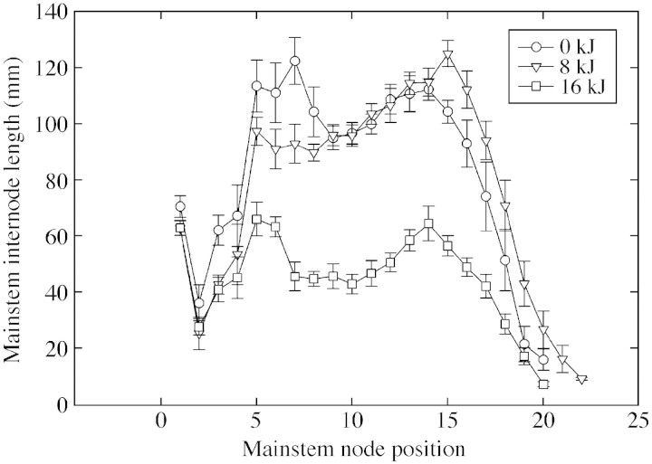
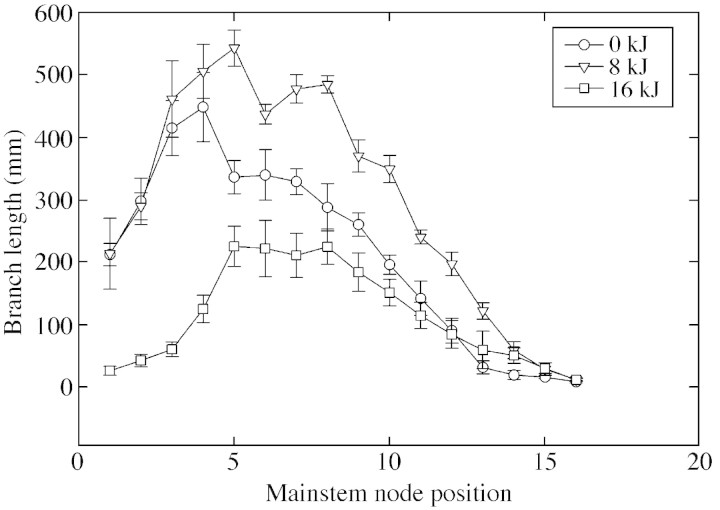
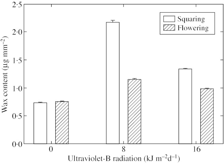
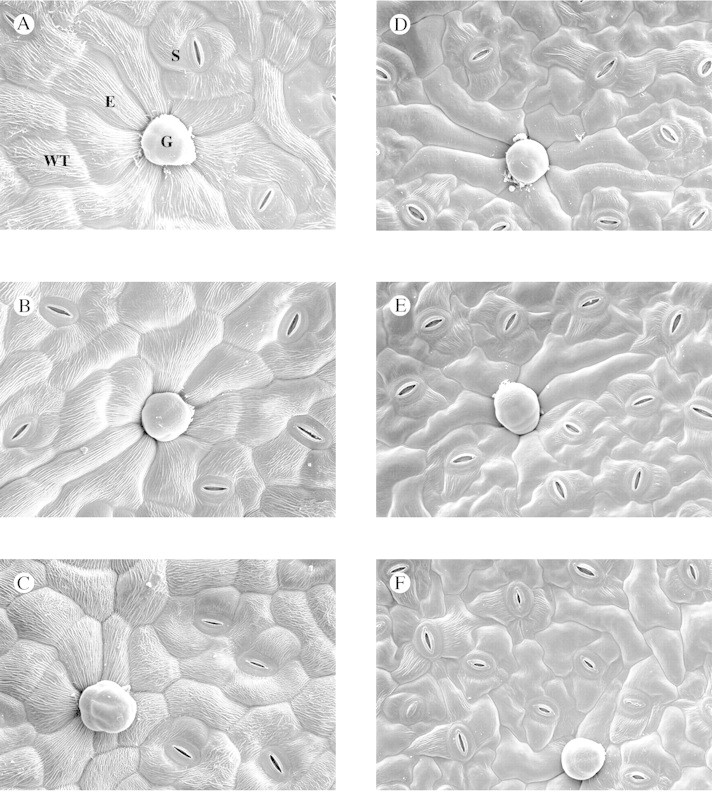
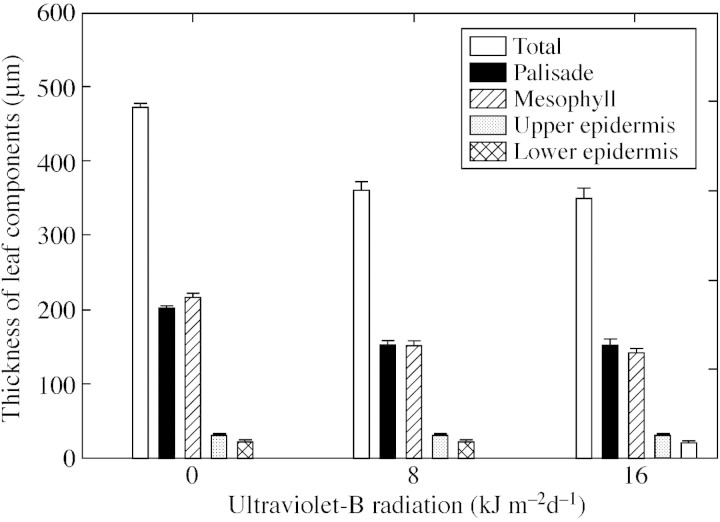
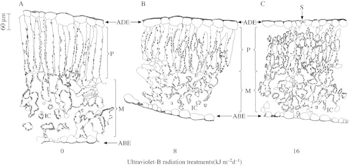
Cotton (Gossypium hirsutum L.) crop, cultivated between 40 degrees N and 40 degrees S, is currently experiencing 2-11 kJ m-2 d-1 of UV-B radiation. This is predicted to increase in the near future. An experiment was conducted to study the effect of enhanced UV-B radiation on vegetative and reproductive morphology and leaf anatomy of cotton in sunlit, controlled environment chambers. From emergence to harvest, cotton plants were exposed to 0, 8 or 16 kJ m-2 d-1 of UV-B in a square wave approach for 8 h from 0800 to 1600 h. Changes in plant height, internode and branch length, mainstem node number, leaf area, length and area of petals and bracts, and anther number per flower were recorded. Epidermal cell and stomatal density, stomatal index, leaf thickness, and epidermal, palisade and mesophyll tissue thickness were also measured. Initial chlorotic symptoms on leaves turned into necrotic patches on continued exposure to enhanced UV-B. Exposure to high UV-B reduced both vegetative and reproductive parameters and resulted in a smaller canopy indicating sensitivity of cotton to UV-B radiation. Enhanced UV-B radiation increased epicuticular wax content on adaxial leaf surfaces, and stomatal index on both adaxial and abaxial leaf surfaces. Leaf thickness was reduced following exposure to UV-B owing to a decrease in thickness of both the palisade and mesophyll tissue, while the epidermal thickness remained unchanged. The vegetative parameters studied were affected only by high levels of UV-B (16 kJ m-2 d-1), whereas the reproductive parameters were reduced at both ambient (8 kJ m-2 d-1) and high UV-B levels. The study shows that cotton plants are sensitive to UV-B at both the whole plant and anatomical level.
Figures
Similar articles
-
Interactive effects of ultraviolet-B radiation and temperature on cotton physiology, growth, development and hyperspectral reflectance.Photochem Photobiol. 2004 May;79(5):416-27. doi: 10.1562/2003-11-19-ra.1. Photochem Photobiol. 2004. PMID: 15191050
-
Leaf and canopy photosynthetic characteristics of cotton (Gossypium hirsutum) under elevated CO2 concentration and UV-B radiation.J Plant Physiol. 2004 May;161(5):581-90. doi: 10.1078/0176-1617-01229. J Plant Physiol. 2004. PMID: 15202715
-
Modelling the structural response of cotton plants to mepiquat chloride and population density.Ann Bot. 2014 Sep;114(4):877-87. doi: 10.1093/aob/mct309. Epub 2014 Jan 31. Ann Bot. 2014. PMID: 24489020 Free PMC article.
-
Developmental reprogramming by UV-B radiation in plants.Plant Sci. 2017 Nov;264:96-101. doi: 10.1016/j.plantsci.2017.09.006. Epub 2017 Sep 13. Plant Sci. 2017. PMID: 28969807 Review.
-
How do UV photomorphogenic responses confer water stress tolerance?Photochem Photobiol. 2003 Dec;78(6):529-34. doi: 10.1562/0031-8655(2003)078<0529:hduprc>2.0.co;2. Photochem Photobiol. 2003. PMID: 14743860 Review.
Cited by
-
Physiological and molecular responses of different rose (Rosa hybrida L.) cultivars to elevated ozone levels.Plant Direct. 2023 Jul 20;7(7):e513. doi: 10.1002/pld3.513. eCollection 2023 Jul. Plant Direct. 2023. PMID: 37484545 Free PMC article.
-
UV-B irradiation changes specifically the secondary metabolite profile in broccoli sprouts: induced signaling overlaps with defense response to biotic stressors.Plant Cell Physiol. 2012 Sep;53(9):1546-60. doi: 10.1093/pcp/pcs096. Epub 2012 Jul 5. Plant Cell Physiol. 2012. PMID: 22773681 Free PMC article.
-
Repression of growth regulating factors by the microRNA396 inhibits cell proliferation by UV-B radiation in Arabidopsis leaves.Plant Cell. 2013 Sep;25(9):3570-83. doi: 10.1105/tpc.113.117473. Epub 2013 Sep 27. Plant Cell. 2013. PMID: 24076976 Free PMC article.
-
Impact of ultraviolet-B radiation on early-season morpho-physiological traits of indica and japonica rice genotypes.Front Plant Sci. 2024 Mar 1;15:1369397. doi: 10.3389/fpls.2024.1369397. eCollection 2024. Front Plant Sci. 2024. PMID: 38495369 Free PMC article.
-
Integrated Analysis of Transcriptomic and Proteomics Data Reveals the Induction Effects of Rotenoid Biosynthesis of Mirabilis himalaica Caused by UV-B Radiation.Int J Mol Sci. 2018 Oct 25;19(11):3324. doi: 10.3390/ijms19113324. Int J Mol Sci. 2018. PMID: 30366418 Free PMC article.
References
-
- BarnesPW, Ballare CL, Caldwell MM.1996. Photomorphogenic effects of UV‐B radiation on plants: consequences for light competition. Journal of Plant Physiology 148: 15–20.
-
- BarnesPW, Maggard S, Holman, SR, Vergara BS.1993. Intraspecific variation in sensitivity to UV‐B radiation in rice. Crop Science 33: 1041–1046.
-
- BecwarMR, Moore FD, Burke MJ.1982. Effects of depletion and enhancement of ultraviolet‐B (280–315 nm) radiation on plants grown at 3000 m elevation. Journal of the American Society of Horticultural Science 107: 771–774.
-
- BondadaBR, Oosterhuis DM, Murphy JB, Kim KS.1996. Effect of water stress on the epicuticular wax composition and ultrastructure of cotton (Gossypium hirsutum L.) leaf, bract, and boll. Environmental and Experimental Botany 36: 61–69.
-
- BondadaBR, Oosterhuis DM, Wullschleger SD, Kim KS, Harris WM.1994. Anatomical considerations related to photosynthesis in cotton leaves, bracts, and the capsule wall. Journal of Experimental Botany 36: 111–118.
