. 2003 Jun;84(6):4023-32.
doi: 10.1016/S0006-3495(03)75128-5.

Selective cell targeting with light-absorbing microparticles and nanoparticles

Affiliations

Selective cell targeting with light-absorbing microparticles and nanoparticles

Costas M Pitsillides et al. Biophys J. 2003 Jun.

Abstract

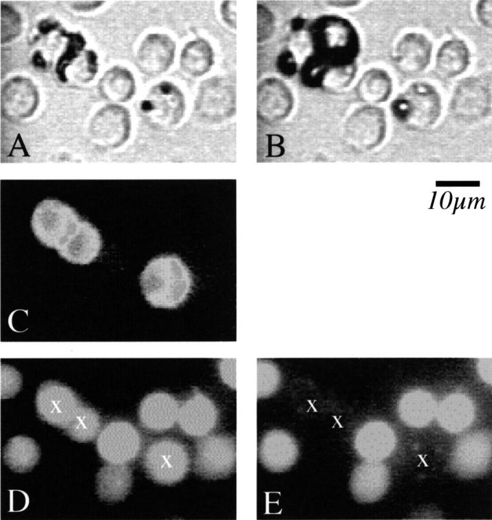
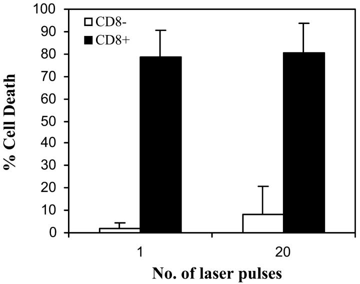
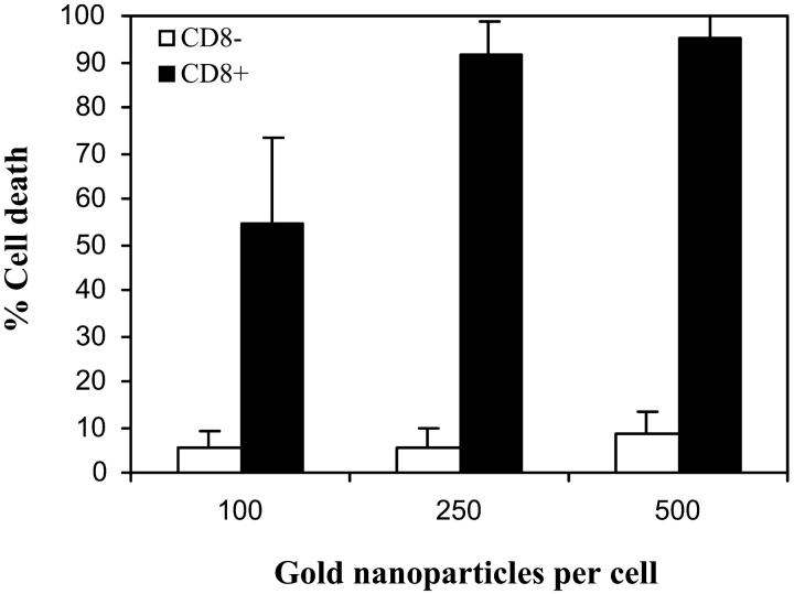
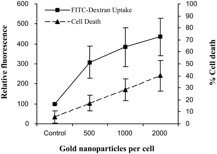
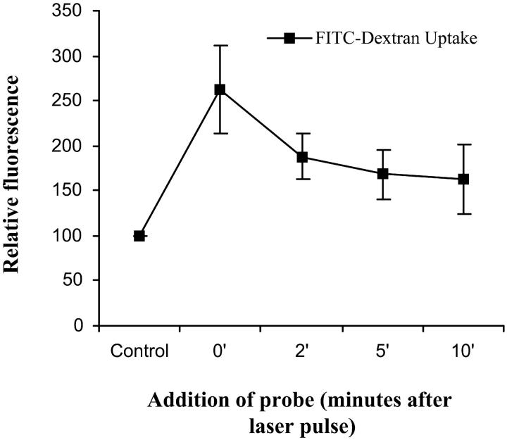
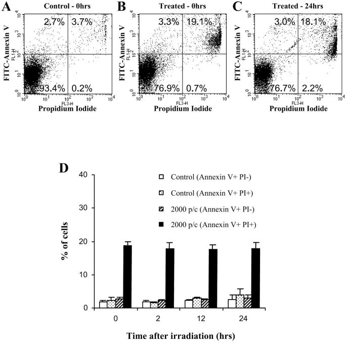
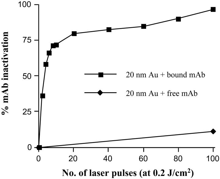
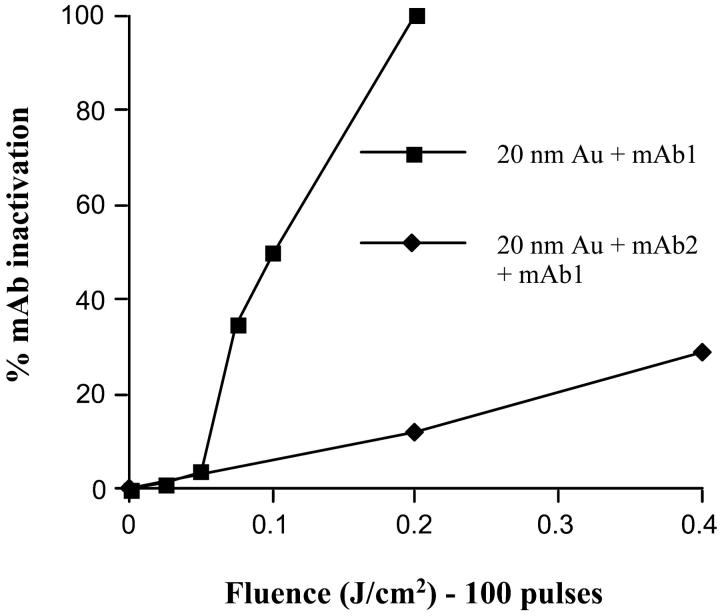
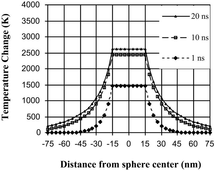
We describe a new method for selective cell targeting based on the use of light-absorbing microparticles and nanoparticles that are heated by short laser pulses to create highly localized cell damage. The method is closely related to chromophore-assisted laser inactivation and photodynamic therapy, but is driven solely by light absorption, without the need for photochemical intermediates (particularly singlet oxygen). The mechanism of light-particle interaction was investigated by nanosecond time-resolved microscopy and by thermal modeling. The extent of light-induced damage was investigated by cell lethality, by cell membrane permeability, and by protein inactivation. Strong particle size dependence was found for these interactions. A technique based on light to target endogenous particles is already being exploited to treat pigmented cells in dermatology and ophthalmology. With exogenous particles, phamacokinetics and biodistribution studies are needed before the method can be evaluated against photodynamic therapy for cancer treatment. However, particles are unique, unlike photosensitizers, in that they can remain stable and inert in cells for extended periods. Thus they may be particularly useful for prelabeling cells in engineered tissue before implantation. Subsequent irradiation with laser pulses will allow control of the implanted cells (inactivation or modulation) in a noninvasive manner.


Figures

FIGURE 1
FIGURE 1
Lymphocytes labeled with anti-CD8 mAb-coated 0.83-μm iron oxide microspheres (5 particles/cell) and double-labeled with anti-CD8 phycoerythrin fluorescent probe. (A) is a bright field image. Iron oxide particles are visible on a subpopulation of the lymphocytes. (B) is a time-resolved image taken ∼100 ns after irradiation with a 565-nm, 20-ns laser pulse at a fluence of 0.35 J/cm2. Rapid growth of microbubbles can be seen around the particles. (C) is a fluorescence image which identifies CD8+ T cells (labeled with PE-conjugated anti-CD8 mAb). (D) and (E) are images of calcein-AM fluorescence before (D) and 1 h (E) after irradiation. Loss of fluorescence by CD8+ lymphocytes indicates loss of viability.
FIGURE 2
FIGURE 2
Viability of T lymphocytes labeled with anti-CD8 Ab + 0.83-μm iron oxide microspheres (5 particles/cell) after irradiation with 1 or 20 pulses (20-ns, 565-nm pulses at 0.35 J/ cm2). Viability assayed 1 h after irradiation using calcein-AM fluorescence.
FIGURE 3
FIGURE 3
Viability of T lymphocytes labeled with 30-nm anti-CD8 immunogold particles, irradiated with 100 laser pulses (565 nm, 20 ns) at 0.5 J/cm2. Calcein-AM fluorescence was used to assay viability 1 h after irradiation.
FIGURE 4
FIGURE 4
(Top) Increase in cell membrane permeability, assayed by FITC-dextran fluorescence uptake, of lymphocytes labeled with 20-nm anti-CD45 immunogold particles and irradiated with 20-ns laser pulses (λ = 532 nm) at 0.5 J/cm2. (bottom) Adding the membrane permeability probe at different time points after irradiation indicates a rapid decline in probe uptake within 2 min.
FIGURE 4
FIGURE 4
(Top) Increase in cell membrane permeability, assayed by FITC-dextran fluorescence uptake, of lymphocytes labeled with 20-nm anti-CD45 immunogold particles and irradiated with 20-ns laser pulses (λ = 532 nm) at 0.5 J/cm2. (bottom) Adding the membrane permeability probe at different time points after irradiation indicates a rapid decline in probe uptake within 2 min.
FIGURE 5
FIGURE 5
Cell death as a function of time after short pulse laser treatment of CD45+ lymphocytes labeled with 20-nm Au particles (2000 per cell) and irradiated with 20-ns laser pulses (λ = 532 nm) at 0.5 J/cm2. Cells were analyzed by apoptosis/necrosis kit (annexin V/propidium iodide). Flow cytometry results (A) untreated cells, (B) immediately after irradiation, and (C) at 24 h after irradiation. The fraction of PI+/Annexin V+ cells (dead cell) remain constant during the 24-h period after irradiation (D). There was also no significant increase in the number of PI/Annexin V+ cells (apoptotic cells) during this period.
FIGURE 6
FIGURE 6
Inactivation of anti-fluorescein rabbit IgG conjugated to 20-nm gold particles. (▪) represent results for inactivation of antibody when it is bound to the gold. (♦) are the results for antibody inactivation when the protein and gold are uncoupled.
FIGURE 7
FIGURE 7
Inactivation of anti-fluorescein rabbit IgG—effect of distance of target from the gold particle absorber. (▪) represent results for inactivation of the primary antibody (mAb1) when it is directly coupled to gold, whereas (♦) are the results for primary antibody inactivation when it is coupled to gold through a secondary antibody (mAb2).
FIGURE 8
FIGURE 8
Theoretical temperature rise in a 30-nm gold particle (Qabs = 2) irradiated with a 20-ns, 532-nm laser pulse at a fluence of 0.5 J/cm2.

Similar articles

Cited by

References

    1. Anderson, R. R. 2000. Lasers in dermatology—a critical update. J. Dermatol. 27:700–705. - PubMed
    1. Anderson, R. R., R. J. Margolis, S. Watenabe, T. Flotte, G. J. Hruza, and J. S. Dover. 1989. Selective photothermolysis of cutaneous pigmentation by Q-switched Nd:YAG laser pulses at 1064, 532, and 355 nm. J. Invest. Dermatol. 93:28–32. - PubMed
    1. Anderson, R. R., and J. A. Parrish. 1983. Selective photothermolysis: precise microsurgery by selective absorption of pulsed radiation. Science. 220:524–527. - PubMed
    1. Beck, S., T. Sakurai, B. K. Eustace, G. Beste, R. Schier, F. Rudert, and D. G. Jay. 2002. Fluorophore-assisted light inactivation: a high throughput tool for direct target validation of proteins. Proteomics. 2:247–255. - PubMed
    1. Brinkmann, R., G. Huettmann, J. Roegener, J. Roider, R. Birngruber, and C. P. Lin. 2000. Origin of retinal pigment epithelium cell damage by pulsed laser irradiance in the nanosecond to microsecond time regimen. Lasers Surg. Med. 27:451–464. - PubMed

Publication types

MeSH terms

Substances

LinkOut - more resources