Structural-functional relationships of the dynein, spokes, and central-pair projections predicted from an analysis of the forces acting within a flagellum
- PMID: 12770914
- PMCID: PMC1302990
- DOI: 10.1016/S0006-3495(03)75136-4
Structural-functional relationships of the dynein, spokes, and central-pair projections predicted from an analysis of the forces acting within a flagellum
Abstract
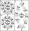
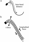
In the axoneme of eukaryotic flagella the dynein motor proteins form crossbridges between the outer doublet microtubules. These motor proteins generate force that accumulates as linear tension, or compression, on the doublets. When tension or compression is present on a curved microtubule, a force per unit length develops in the plane of bending and is transverse to the long axis of the microtubule. This transverse force (t-force) is evaluated here using available experimental evidence from sea urchin sperm and bull sperm. At or near the switch point for beat reversal, the t-force is in the range of 0.25-1.0 nN/ micro m, with 0.5 nN/ micro m the most likely value. This is the case in both beating and arrested bull sperm and in beating sea urchin sperm. The total force that can be generated (or resisted) by all the dyneins on one micron of outer doublet is also approximately 0.5 nN. The equivalence of the maximum dynein force/ micro m and t-force/ micro m at the switch point may have important consequences. Firstly, the t-force acting on the doublets near the switch point of the flagellar beat is sufficiently strong that it could terminate the action of the dyneins directly by strongly favoring the detached state and precipitating a cascade of detachment from the adjacent doublet. Secondly, after dynein release occurs, the radial spokes and central-pair apparatus are the structures that must carry the t-force. The spokes attached to the central-pair projections will bear most of the load. The central-pair projections are well-positioned for this role, and they are suitably configured to regulate the amount of axoneme distortion that occurs during switching. However, to fulfill this role without preventing flagellar bend formation, moveable attachments that behave like processive motor proteins must mediate the attachment between the spoke heads and the central-pair structure.
Figures


Similar articles
-
Testing the geometric clutch hypothesis.Biol Cell. 2004 Dec;96(9):681-90. doi: 10.1016/j.biolcel.2004.08.001. Biol Cell. 2004. PMID: 15567522 Review.
-
Dynein arms are oscillating force generators.Nature. 1998 Jun 18;393(6686):711-4. doi: 10.1038/31520. Nature. 1998. PMID: 9641685
-
The geometric clutch as a working hypothesis for future research on cilia and flagella.Ann N Y Acad Sci. 2007 Apr;1101:477-93. doi: 10.1196/annals.1389.024. Epub 2007 Feb 15. Ann N Y Acad Sci. 2007. PMID: 17303832 Review.
-
Measurement of the force produced by an intact bull sperm flagellum in isometric arrest and estimation of the dynein stall force.Biophys J. 2000 Jul;79(1):468-78. doi: 10.1016/S0006-3495(00)76308-9. Biophys J. 2000. PMID: 10866972 Free PMC article.
-
Mechanical properties of the passive sea urchin sperm flagellum.Cell Motil Cytoskeleton. 2009 Sep;66(9):721-35. doi: 10.1002/cm.20401. Cell Motil Cytoskeleton. 2009. PMID: 19536829
Cited by
-
Diurnal Variations in the Motility of Populations of Biflagellate Microalgae.Biophys J. 2020 Nov 17;119(10):2055-2062. doi: 10.1016/j.bpj.2020.10.006. Epub 2020 Oct 20. Biophys J. 2020. PMID: 33091375 Free PMC article.
-
Oscillatory movement of a dynein-microtubule complex crosslinked with DNA origami.Elife. 2022 Jun 24;11:e76357. doi: 10.7554/eLife.76357. Elife. 2022. PMID: 35749159 Free PMC article.
-
Push-me-pull-you: how microtubules organize the cell interior.Eur Biophys J. 2008 Sep;37(7):1271-8. doi: 10.1007/s00249-008-0321-0. Epub 2008 Apr 11. Eur Biophys J. 2008. PMID: 18404264 Free PMC article. Review.
-
A computational model of dynein activation patterns that can explain nodal cilia rotation.Biophys J. 2015 Jul 7;109(1):35-48. doi: 10.1016/j.bpj.2015.05.027. Biophys J. 2015. PMID: 26153700 Free PMC article.
-
FAP206 is a microtubule-docking adapter for ciliary radial spoke 2 and dynein c.Mol Biol Cell. 2015 Feb 15;26(4):696-710. doi: 10.1091/mbc.E14-11-1506. Epub 2014 Dec 24. Mol Biol Cell. 2015. PMID: 25540426 Free PMC article.
References
-
- Ashkin, A., K. Shultze, J. M. Dziedzic, U. Euteneur, and M. Schliwa. 1990. Force generation of organelle transport measured in vivo by an infrared laser trap. Nature. 348:346–348. - PubMed
-
- Brokaw, C. J. 1965. Non-sinusoidal bending waves of sperm flagella. J. Exp. Biol. 43:155–169. - PubMed
-
- Brokaw, C. J. 1984. Automated methods for estimation of sperm flagellar bending parameters. Cell Motil. 4:417–430. - PubMed
-
- Brokaw, C. J. 1990. The sea urchin spermatozoon. Bioessays. 12:449–452. - PubMed
Publication types
MeSH terms
Substances
LinkOut - more resources
Full Text Sources
Miscellaneous

