. 2003 May;139(2):219-31.
doi: 10.1038/sj.bjp.0705216.

Thymidine phosphorylase induces angiogenesis in vivo and in vitro: an evaluation of possible mechanisms

Affiliations

Thymidine phosphorylase induces angiogenesis in vivo and in vitro: an evaluation of possible mechanisms

Shiladitya Sengupta et al. Br J Pharmacol. 2003 May.

Abstract

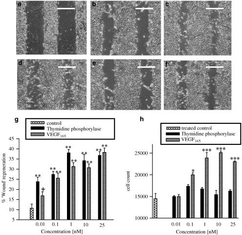
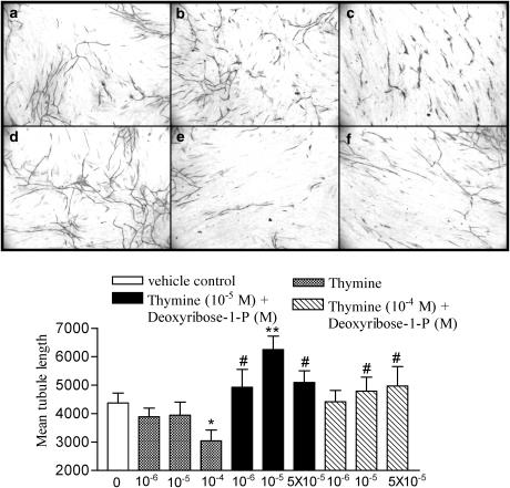
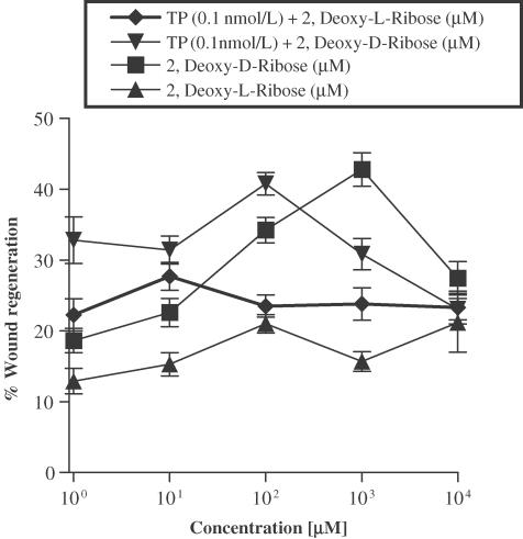
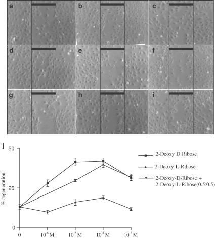
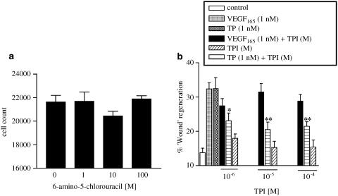
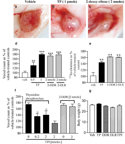
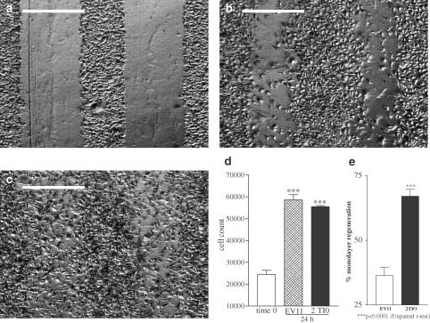
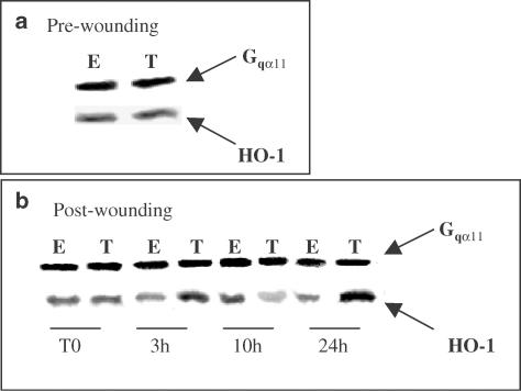
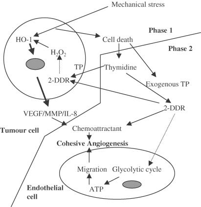
1 Thymidine phosphorylase (TP) is elevated in the plasma of cancer patients, and has been implicated in pathophysiological angiogenesis. However, the downstream signals underlying this implication remain obscure. The purpose of the present study was to examine the effects of TP on the neovascularisation response in vitro and in vivo. 2 Both TP and its catalytic product, 2-deoxy-D-ribose-1-phosphate, and downstream 2-deoxy-D-ribose (2-DDR) promoted endothelial tubulogenesis in vitro, and the regeneration of a wounded monolayer of endothelial cells without exerting any mitogenic effect. In vivo, both TP and 2-DDR promoted the development of functional vasculature into an avascular sponge. A TP inhibitor, 6-amino-5-chlorouracil, was able to partially reverse the effects of TP, but had no effect on the 2-DDR-induced angiogenesis. 3 Enhanced monolayer regeneration was observed with TP-cDNA-transfected bladder carcinoma cells. The transfection of TP-cDNA, however, did not confer any proliferative advantage. The regeneration of TP overexpressing cells was associated with a time-dependent expression of the enzyme haeme-oxygenase (HO-1). 4 The present study demonstrates that both TP and its ribose-sugar metabolites induce angiogenesis by mediating a cohesive interplay between carcinoma and endothelial cells. The induction of HO-1 in TP-transfected cells suggests that it could be a possible downstream signal for the angiogenic effects of TP. Furthermore, reducing sugars have been shown to induce oxidative stress, and ribose could be a possible cause for the upregulation of HO-1, which has been implicated in the release of angiogenic factors. Therefore, we postulate that 2-DDR could be mediating the angiogenic effects of TP possibly through an oxidative stress mechanism and additionally getting integrated in the endothelial metabolic machinery.


Figures

Figure 1
Figure 1
The effect of thymidine phosphorylase and VEGF165 on HUVECs monolayer regeneration, following a mechanical injury. The top panel of phase contrast micrographs show: (a) the total denudation at the time of injury (T0); (b) basal regrowth in the presence of vehicle (5% FCS); (c) regeneration in the presence of VEGF (1 nM); (d) regeneration in the presence of thymidine phosphorylase (TP) (1 nM); (e) preincubation for 1 h and the continuous presence of TP inhibitor, reverts the TP-induced regeneration to basal level; (f) TP inhibitor has no effect on the VEGF165-induced recovery. Images were captured with a Nikon Diaphot inverted microscope at × 4 objective, coupled to a CCD (JVC), and grabbed using a Q500 Leica software. The calibration bar represents 400 μm. The graphs show the concentration–effect curve of TP and VEGF165, on (g) the recovery of a ‘wounded' area, and (h) the proliferation of endothelial cell. A synchronised monolayer of HUVECs was injured using a multichannel wounder, producing 11 linear lesions, and transferred to fresh media supplemented with 5% FCS, with appropriate treatments. They were incubated for a further 24 h, following which, they were either fixed and image analysed, or trypsinised and counted using a haemocytometer. Data expressed are mean±s.e.m. of at least four separate experiments with quadruplicate wells in each. In the wound recovery experiment, data are shown as percentage of T0 values. *P<0.05, **P<0.01, ***P<0.001 vs vehicle-treated controls, +P<0.05 vs corresponding TP-treated group.
Figure 2
Figure 2
Effect of 2-deoxy-D-ribose-1-phosphate (2-DDR-1-P) and thymine on tube formation by endothelial cells in a coculture system. Photomicrographs depict: (a) vehicle-treated cells, (b) VEGF-induced tubulogenesis, (c) suramin-induced inhibition of tubulogenesis, (d) tube formation induced by 2-DDR-1-P (10−6 M), (e) inhibition of tubulogenesis by thymine (10−4 M), and (f) reversal of thymine-inhibition by 2-DDR-1-P (10−4 M). A coculture of endothelial cells and fibroblasts (Angiokit) was used to study the tube formation by endothelial cells. The AngioKit was seeded with cells on day 0, and the optimised growth medium was changed on days 3, 5, 7, 10 and 12. It was then fixed, and stained for CD31, on day 14. Suramin (20 μM) and VEGF (2 ng ml−1) were used as negative and positive controls, respectively. Thymine and 2-DDR-1-P were added on day 4. The appropriate treatments were replenished with each medium change. Graph (g) shows the comparison of venule length following different treatments, as measured using a ‘AngioSys' image analysis system. Four images were grabbed per well, from the four quadrants. Experiments were run in quadruplicate, and data were expressed as the mean±s.e.m. *P<0.05, **P<0.01 vs vehicle control; #P<0.05 vs matched thymine concentration.
Figure 3
Figure 3
Effect of 2-deoxy-D-ribose and 2-deoxy-L-ribose on the proliferation of endothelial cell, and regeneration following a mechanical injury. Cells were grown and injured as described in Methods, and transferred to fresh media supplemented with 5% FCS, with and without the sugars. They were incubated for a further 24 h following which, they were either fixed and image analysed, or trypsinised and counted using a haemocytometer. Nomarsky images were captured using a Nikon Diaphot ( × 4 objective) as described in Methods. Photomicrographs depict (a) the total wounded area at T0, and the recovery in (b) vehicle-treated cells, (c) 2-deoxy-D-ribose (10−5 M)-treated cells and (d) 2-deoxy-L-ribose (10−5 M)-treated cells, 24 h postinjury. The graphs show the concentration–effect curves depicting (e) wound recovery and (f) cell count, following treatment with increasing concentrations of the sugars. Data expressed are mean±s.e.m. of at least four separate experiments with duplicate/quadruplicate wells in each. In the wound recovery experiment, data are shown as percentage of 0 h values. *P<0.001, #P<0.05 vs vehicle-treated control. The bar represents 400 μm.
Figure 4
Figure 4
Effect of thymidine phosphorylase, on the 2-deoxy-D-ribose- and 2-deoxy-L-ribose-induced regeneration of the monolayer, postinjury. Cells were plated at a density of 20,000 cells/well, in a 24-well plate containing a coverslip in each well. They were cultured to confluence in 20% FCS-supplemented media and then transferred to media with 1% FCS for 24 h. The monolayer was then lesioned using a multichannel wounder (described in Methods), and transferred to fresh media supplemented with 5% FCS, with and without the sugars. They were incubated for a further 24 h, following which, they were fixed and image-analysed. Data expressed are mean±s.e.m. of at least four separate experiments with quadruplicate wells in each. Data are shown as percentage of 0 h values.
Figure. 5
Figure. 5
Effect of combination of 2-deoxy-D-ribose and 2-deoxy-L-ribose in a 1 : 1 ratio on the regeneration of a injured monolayer of endothelial cells. HUVECs were grown to confluence in endothelial growth medium (Clonetics, U.S.A.), and synchronised in 0.5% serum. The confluent monolayer was injured as described in the Methods section, and allowed to recover in EGM supplemented with 5% fetal bovine serum for 24 h in the presence of different concentrations of ribose sugars and their combinations. The cells were fixed at 24 h, and image analysed using an Olympus inverted microscope using a × 10 objective. The photomicrographs depict the total recovery following treatment with (a) vehicle, (b) 2-DDR (10−6 M), (c) 2-DDR (10−5 M), (d) 2-DDR (10−4 M), (e) 2-DLR (10−6 M), (f) 2-DLR (10−5 M), (g) 2-DLR (10−4 M), (h) 2-DDR+2-DLR (10−5 M), (I) 2-DDR+2-DLR (10−4 M). The graph (j) represents the total recovery at 24 h, expressed as a percentage of the initial area denuded. Data represents mean±s.e.m. from three separate experiments with duplicate wells in each.
Figure 6
Figure 6
Effect of a thymidine phosphorylase inhibitor (TP), 6-amino-5-chlorouracil, on (a) the proliferation of endothelial cell, and on (b) the thymidine phosphorylase- and VEGF165-induced regeneration of the monolayer, postinjury. Cells were plated at a density of 20,000 cells well−1, in a 24-well plate containing a coverslip in each well. They were cultured to confluence in 20% FCS-supplemented media and then transferred to media with 1% FCS for 24 h. The TP inhibitor was added 1 h prior to ‘wounding'. The monolayer was then lesioned using a multichannel wounder, and transferred to fresh media supplemented with 5% FCS, with and without the compounds. They were incubated for a further 24 h, following which, they were either fixed and image analysed, or trypsinised and counted using a haemocytometer. Data expressed are mean±s.e.m. of at least three separate experiments with duplicate/quadruplicate wells in each. In the wound recovery experiment, data are shown as percentage of T0 values. *P<0.05, **P<0.01 vs thymidine phosphorylase-treated controls.
Figure 7
Figure 7
Effect of thymidine phosphorylase (TP) and sugars, 2 deoxy-D-ribose (2-DDR) and deoxy-L-ribose, in a sponge granuloma model of angiogenesis. Pictomicrographs depict (a) neovascularisation in a vehicle-treated sponge, (b) TP (1 pmol/sponge for 10 days)-treated sponge increased the angiogenic response, and (c) a 2-deoxy-D-ribose (2 nmol/sponge × 10 days)-treated sponge implant. The graphs show the angiogenesis in the sponge implant, as quantified by (d) vessel counts, and (e) measuring the total 6 min 133Xe-clearance, indicating that the treatment with TP, 2-DDR and 2-DLR promoted a strong angiogenic response. Graph (f) shows the effects of a thymidine phosphorylase inhibitor on the angiogenesis induced by TP, and 2-DDR in the sponge granuloma model of angiogenesis, while (g) depicts the body weight of the mice following different treatments. Results were calculated from a minimum of two experiments with at least three–four replicates per test group. Data are expressed as mean±s.e.m. *P<0.05, **P<0.01 and ***P<0.001 vs vehicle-treated controls. +P<0.05 vs TP-treated controls.
Figure 8
Figure 8
Response of TP-transfected and wild-type human bladder carcinoma cells to mechanical denudation of the confluent monolayer. The cell line RT112 was transfected with human TP cDNA to generate the 2T10 cell line. EV11 was generated as the empty vector control transfectant. The cells were grown to confluence on Thermanox coverslips in medium supplemented with 10% FCS and glutamine. They were then wounded using a multichannel mechanical wounder, and transferred to fresh medium. They were incubated for a further 24 h, after which they were either fixed and image-analysed, or trypsinised and counted using a haemocytometer. Nomarsky images were captured using a Nikon Diaphot ( × 4 objective) as described in Methods. Photomicrographs depicting (a) a wounded area at time 0 where the cells were fixed immediately after wounding, wound recovery in (b) regeneration by EV11 cells, (c) regrowth of 2T10 cells, 24 h postinjury. Graphs depict (e) cell count and (f) wound recovery, following incubation for 24 h post-injury. Data expressed are mean±s.e.m. of at least three separate experiments with duplicate wells in each. In the wound recovery experiment, data are shown as percentage of 0 h values. ***P<0.001.
Figure 9
Figure 9
Level of hemeoxygenase 1 (HO-1) at different time points following mechanical injury to a confluent monolayer of human bladder carcinoma cells. The cell line RT112 was transfected with human TP cDNA to generate the 2T10 (T) cell line. EV11 (E) was generated as the empty vector control transfectant. Cells were grown to confluence on Thermanox coverslips, and then wounded using a multichannel mechanical wounder, and transferred to fresh medium. At fixed time intervals, the cells were lysed in buffer and samples were separated on a 10% SDS–PAGE. Haemeoxygenase was detected using a goat polyclonal antibody raised against a peptide mapping at the carboxy terminus of HO-1 of human origin. A concomitant control for Gαq11was run to establish the uniformity of protein loading. Block (a) shows the levels of HO-1 prior to the induction of the injury, while (b) shows the time course of the expression of HO-1 in the cells after injury.
Figure 10
Figure 10
A proposed mechanism for the angiogenic effects of thymidine phosphorylase. Mechanical stress can lead to cell death (normal inside a solid tumour), and result in the release of thymidine in the microenvironment. Thymidine can enter live carcinoma cells, and is broken down by TP to 2-DDR-1-P, which can be dephosphorylated to 2-DDR. Inside a tumour cell (phase 1), reducing sugars can undergo rearrangement reactions, leading to the generation of free radicals. The concomitant upregulation of HO-1 has been implicated in increasing the expression of VEGF, MMPs and IL-8. These can then act on the host endothelial cells to induce an angiogenic effect in vivo. Furthermore, CO released would stabilise the neo-vessels through an antiapoptotic effect. Additionally, as seen in phase 2, the TP released from injured cells, or added exogenously, can act on thymidine and lead to the generation of 2-DDR, which is a chemotactic/chemokinetic factor, and promote angiogenesis. The 2-DDR that enters the cell can also be incorporated into the glycolytic machinery, and generate energy that can further serve towards a migratory phenotype. This model explains why both 2-DDR and 2-DLR could exert angiogenic effects in vivo, but only 2-DDR was active in vitro. Thin arrows indicate possible links elucidated in this study, while hashed arrows are putative links. The thicker arrows indicate pathways established by other studies (Brown et al., 2000).

Similar articles

Cited by

References

    1. BOYLE J.J., WILSON B., BICKNELL R., HARROWER S., WEISSBERG P.L., FAN T.P. Expression of angiogenic factor thymidine phosphorylase and angiogenesis in human atherosclerosis. J. Pathol. 2000;192:234–242. - PubMed
    1. BROWN N.S., BICKNELL R.Thymidine phosphorylase, 2-deoxy-D-ribose and angiogenesis Biochem. J. 19983341–8.(Part 1) - PMC - PubMed
    1. BROWN N.S., JONES A., FUJIYAMA C., HARRIS A.L., BICKNELL R. Thymidine phosphorylase induces carcinoma cell oxidative stress and promotes secretion of angiogenic factors. Cancer Res. 2000;60:6298–6302. - PubMed
    1. CHOI A.M., ALAM J. Heme oxygenase-1: function, regulation, and implication of a novel stress-inducible protein in oxidant-induced lung injury. Am. J. Respir. Cell Mol. Biol. 1996;15:9–19. - PubMed
    1. CURRIE M.J., GUNNINGHAM S.P., HAN C., SCOTT P.A., ROBINSON B.A., HARRIS A.L., FOX S.B. Angiopoietin-1 is inversely related to thymidine phosphorylase expression in human breast cancer, indicating a role in vascular remodeling. Clin. Cancer Res. 2001;7:918–927. - PubMed

Publication types

MeSH terms

LinkOut - more resources