

Mechanisms involved in the stimulation of prostacyclin synthesis by human lymphocytes in human umbilical vein endothelial cells
- PMID: 12770937
- PMCID: PMC1573851
- DOI: 10.1038/sj.bjp.0705253
Mechanisms involved in the stimulation of prostacyclin synthesis by human lymphocytes in human umbilical vein endothelial cells
Abstract
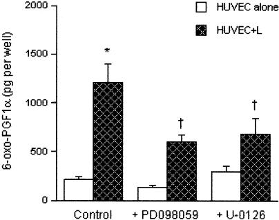
1 Endothelial cells play an important role in the modulation of vascular tone because of their ability to produce vasoactive substances such as prostacyclin (PGI(2)). Cell-cell contact between human umbilical vein endothelial cells (HUVEC) and peripheral blood lymphocytes has been shown to stimulate endothelial PGI(2) synthesis by increasing free arachidonic acid availability through endothelial cytosolic phospholipase A2 (cPLA(2)) activation. In this study, we sought to determine whether phospholipase C (PLC) and D (PLD) activation also contributes, besides cPLA(2), to the lymphocyte-induced PGI(2) synthesis in HUVEC, and to delineate further the potential mechanisms of cPLA(2) activation triggered by the interaction of HUVEC with lymphocytes. 2 Pretreatment of endothelial cells with the PI-PLC inhibitor U-73122 before the coincubation with lymphocytes markedly inhibited the PGI(2) output whereas the diacylglycerol (DAG) lipase inhibitor RHC 80267 and ethanol had no effect. These results suggest that PLC may be involved through inositol trisphosphate generation and calcium mobilization, and that neither DAG nor phosphatidic acid (PtdOH) was used as sources of arachidonic acid. 3 The stimulated PGI(2) synthesis was protein kinase C (PKC)-independent but strongly inhibited by the mitogen-activated protein kinase kinase (MEK) inhibitors PD98059 and U-0126 and by the Src kinase inhibitor PP1. 4 Immunoblot experiments showed an increased phosphorylation of the extracellular signal-regulated kinases 1/2 (ERK1/2) upon lymphocyte addition till 4 h coincubation. Phosphorylation was markedly inhibited by U-0126 and PP1 addition. 5 Collectively, these results suggest that the signaling cascade triggered by lymphocytes in endothelial cells involves an Src kinase/ERK1/2 pathway leading to endothelial cPLA(2) activation.
Figures


Similar articles
-
Agonist-specific cross talk between ERKs and p38(mapk) regulates PGI(2) synthesis in endothelium.Am J Physiol Cell Physiol. 2001 Oct;281(4):C1266-76. doi: 10.1152/ajpcell.2001.281.4.C1266. Am J Physiol Cell Physiol. 2001. PMID: 11546664
-
Vascular endothelial growth factor stimulates prostacyclin production and activation of cytosolic phospholipase A2 in endothelial cells via p42/p44 mitogen-activated protein kinase.FEBS Lett. 1997 Dec 22;420(1):28-32. doi: 10.1016/s0014-5793(97)01481-6. FEBS Lett. 1997. PMID: 9450544
-
sPLA(2) cooperates with cPLA(2)alpha to regulate prostacyclin synthesis in human endothelial cells.Biochem Biophys Res Commun. 2001 Oct 5;287(4):881-7. doi: 10.1006/bbrc.2001.5681. Biochem Biophys Res Commun. 2001. PMID: 11573947
-
Phospholipases and phagocytosis: the role of phospholipid-derived second messengers in phagocytosis.Int J Biochem Cell Biol. 1999 Mar-Apr;31(3-4):415-30. doi: 10.1016/s1357-2725(98)00108-3. Int J Biochem Cell Biol. 1999. PMID: 10224668 Review.
-
Regulation of endothelial prostacyclin synthesis by protease-activated receptors: mechanisms and significance.Pharmacol Rep. 2008 Jan-Feb;60(1):109-18. Pharmacol Rep. 2008. PMID: 18276992 Review.
Cited by
-
p38 mitogen-activated protein kinase mediates lipopolysaccharide and tumor necrosis factor alpha induction of shiga toxin 2 sensitivity in human umbilical vein endothelial cells.Infect Immun. 2008 Mar;76(3):1115-21. doi: 10.1128/IAI.01300-07. Epub 2007 Dec 17. Infect Immun. 2008. PMID: 18086809 Free PMC article.
-
Combinatory action of VEGFR2 and MAP kinase pathways maintains endothelial-cell integrity.Cell Res. 2011 Jul;21(7):1080-7. doi: 10.1038/cr.2011.41. Epub 2011 Mar 22. Cell Res. 2011. PMID: 21423276 Free PMC article.
-
Bile acid reflux contributes to development of esophageal adenocarcinoma via activation of phosphatidylinositol-specific phospholipase Cgamma2 and NADPH oxidase NOX5-S.Cancer Res. 2010 Feb 1;70(3):1247-55. doi: 10.1158/0008-5472.CAN-09-2774. Epub 2010 Jan 19. Cancer Res. 2010. PMID: 20086178 Free PMC article.
References
-
- BALSINDE J., DIEZ E., MOLLINEDO F. Arachidonic acid release from diacylglycerol in human neutrophils. Translocation of diacylglycerol-deacylating enzyme activities from an intracellular pool to plasma membrane upon cell activation. J. Biol. Chem. 1991;266:15638–15643. - PubMed
-
- CARROLL M.P., MAY W.S. Protein kinase C-mediated serine phosphorylation directly activates Raf-1 in murine hematopoietic cells. J. Biol. Chem. 1994;269:1249–1256. - PubMed
-
- CLARK J.D., SCHIEVELLA A.R., NALEFSKI E.A., LIN L.L. Cytosolic phospholipase A2. J. Lipid Mediat. Cell. Signal. 1995;12:83–117. - PubMed
-
- COBB M.H. MAP kinase pathways. Progr. Biophys. Mol. Biol. 1999;71:479–500. - PubMed
Publication types
MeSH terms
Substances
LinkOut - more resources
Full Text Sources
Research Materials
Miscellaneous
