

A clinical follow up of PRK and LASIK in eyes with preoperative abnormal corneal topographies
- PMID: 12770960
- PMCID: PMC1771711
- DOI: 10.1136/bjo.87.6.682
A clinical follow up of PRK and LASIK in eyes with preoperative abnormal corneal topographies
Abstract
Aim: To assess the safety and predictability of photorefractive keratotomy (PRK) and laser in situ keratomileusis (LASIK) based on preoperative corneal topography.
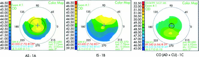
Methods: A non-randomised comparative study was carried out on 84 eyes that presented with topographic abnormalities before undergoing PRK (n = 44) or LASIK (n = 40) procedures. 84 spherical equivalent paired normal eyes served as the control group. Either PRK or LASIK procedures were performed on 168 eyes using the Summit apex plus excimer laser. Topographic abnormalities, including apex displacement (AD), increased asphericity (AS), meridional irregularity (MI), increased inferior-superior asymmetry (IS), increased curvature (CU), and combined features (CO), were assessed preoperatively using the EyeSys analysis system. Safety and predictability of the two procedures were defined as a postoperative visual acuity of 20/40 or better and the loss of one or more lines of spectacle corrected visual acuity (SCVA).
Results: All patients were followed for 6 months. There was a significant loss of best corrected visual acuity in the PRK-AD (p<0.001), PRK-CO (p<0.05), and LASIK-AS (p<0.001) patients. The number of eyes within plus or minus 1.0D of the surgical plan postoperatively was similar in all groups.
Conclusion: These data suggest that although predictability was similar, PRK and LASIK performed in corneas with topographic abnormalities might cause loss of vision.
Figures
References
-
- Johnson JD, Azar DT. Surgically induced topographical abnormalities after LASIK: management of central islands, corneal ectasia, decentration, and irregular astigmatism. Curr Opin Ophthalmol 2001;12:309–17. - PubMed
-
- Holland SP, Srivannaboon S, Reinstein DZ. Avoiding serious corneal complications of laser assisted in situ keratomileusis and photorefractive keratectomy. Ophthalmology 2000;107:640–52. - PubMed
-
- Wilson SE, Klyce SD. Screening for corneal topographic abnormalities before refractive surgery. Ophthalmology 1994;101:147–52. - PubMed
-
- Neves R, Schor P, Nosé W. Ceratocone suspeito em pacientes candidatos a ceratotomia radial. Arquivos Brasileiros de Oftalmologia 1994;57:202–4.
Publication types
MeSH terms
LinkOut - more resources
Full Text Sources
Medical
Research Materials
Miscellaneous