

Comparison of localised nerve fibre layer defects in normal tension glaucoma and primary open angle glaucoma
- PMID: 12770963
- PMCID: PMC1771704
- DOI: 10.1136/bjo.87.6.695
Comparison of localised nerve fibre layer defects in normal tension glaucoma and primary open angle glaucoma
Abstract
Aim: To compare the pattern of localised nerve fibre layer (NFL) defects in normal tension glaucoma (NTG) and primary open angle glaucoma (POAG).
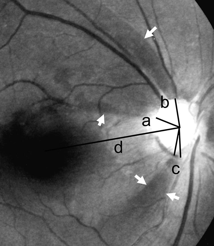
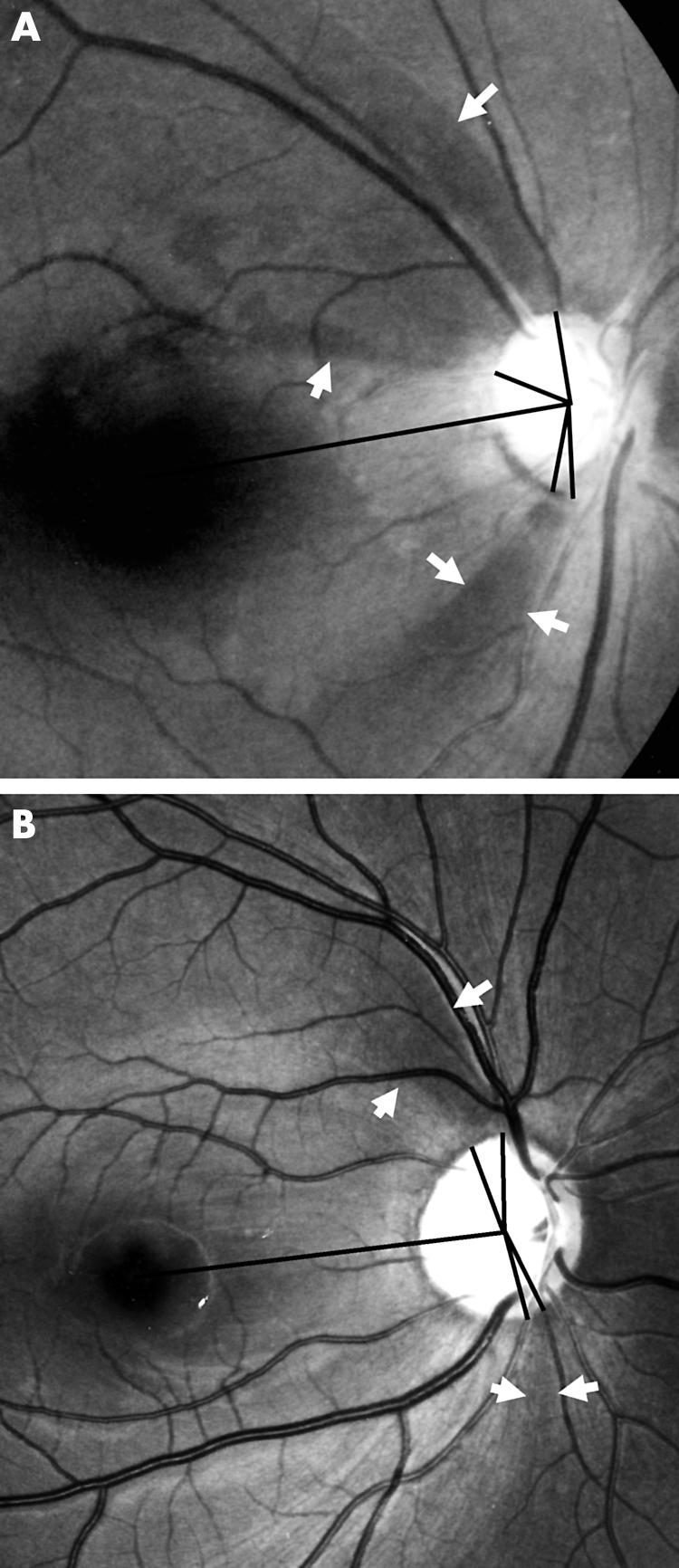
Methods: 50 NTG eyes and 36 POAG eyes, all with localised NFL defects, were enrolled. On retinal NFL photography, the proximity of the defect to the centre of the fovea (angle alpha) and the sum of the angular width of the defects (angle beta) were determined. Angle alpha was the angle made by a line from the centre of the fovea to the disc centre and a line from the disc centre to the disc margin, where the nearest border of the defect met. The patterns of localised NFL defects in NTG and POAG were compared with angles alpha and beta. Independent t test was used for statistical analysis.
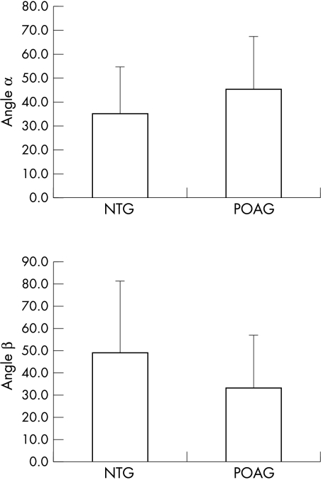
Results: Angle alpha in NTG (35.1 (SD 20.0) degrees ) was significantly smaller than that of POAG (45.9 (21.9) degrees ) (p=0.02), while angle beta in NTG (49.0 (31.9) degrees ) was significantly larger than that of POAG (33.1 (23.9) degrees ) (p=0.01).
Conclusions: The pattern of NFL defects in NTG was different from that in POAG. Localised NFL defects in NTG were closer to the fovea and wider in width than those in POAG.
Figures
Similar articles
-
Localized wedge-shaped defects of retinal nerve fiber layer and disc hemorrhage in glaucoma.Ophthalmology. 1999 Sep;106(9):1762-7. doi: 10.1016/S0161-6420(99)90347-0. Ophthalmology. 1999. PMID: 10485548 Clinical Trial.
-
Comparison of retinal nerve fiber layer and macular thickness for discriminating primary open-angle glaucoma and normal-tension glaucoma using optical coherence tomography.Clin Exp Optom. 2016 Jul;99(4):373-81. doi: 10.1111/cxo.12366. Epub 2016 Mar 21. Clin Exp Optom. 2016. PMID: 26996257
-
[The retinal nerve fiber layer defect and its related clinical features in early primary open-angle glaucoma].Zhonghua Yan Ke Za Zhi. 2001 May;37(3):193-6. Zhonghua Yan Ke Za Zhi. 2001. PMID: 11864420 Chinese.
-
[A challenge to primary open-angle glaucoma including normal-pressure. Clinical problems and their scientific solution].Nippon Ganka Gakkai Zasshi. 2012 Mar;116(3):233-67; discussion 268. Nippon Ganka Gakkai Zasshi. 2012. PMID: 22568103 Review. Japanese.
-
[Open-angle glaucoma clinical presentation and management].Nippon Ganka Gakkai Zasshi. 2001 Dec;105(12):828-42. Nippon Ganka Gakkai Zasshi. 2001. PMID: 11802456 Review. Japanese.
Cited by
-
Common variants on chromosome 9p21 are associated with normal tension glaucoma.PLoS One. 2012;7(7):e40107. doi: 10.1371/journal.pone.0040107. Epub 2012 Jul 5. PLoS One. 2012. PMID: 22792221 Free PMC article.
-
Central retinal vascular trunk deviation in unilateral normal-tension glaucoma.PLoS One. 2021 Jul 20;16(7):e0254889. doi: 10.1371/journal.pone.0254889. eCollection 2021. PLoS One. 2021. PMID: 34283884 Free PMC article.
-
Angular Location of Retinal Nerve Fiber Layer Defect: Association With Myopia and Open-Angle Glaucoma.Invest Ophthalmol Vis Sci. 2020 Sep 1;61(11):13. doi: 10.1167/iovs.61.11.13. Invest Ophthalmol Vis Sci. 2020. PMID: 32902578 Free PMC article.
-
Defective angles of localized retinal nerve fiber layer reflect the severity of visual field defect- a cross-sectional analysis.BMC Ophthalmol. 2020 Apr 9;20(1):141. doi: 10.1186/s12886-020-01396-y. BMC Ophthalmol. 2020. PMID: 32272929 Free PMC article.
-
Racial Differences in Diagnostic Accuracy of Retinal Nerve Fiber Layer Thickness in Primary Open-Angle Glaucoma.Am J Ophthalmol. 2024 Mar;259:7-14. doi: 10.1016/j.ajo.2023.10.012. Epub 2023 Oct 29. Am J Ophthalmol. 2024. PMID: 38708401 Free PMC article.
References
-
- Caprioli J, Spaeth GL. Comparison of visual field defects in the low-tension glaucomas with those in the high-tension glaucomas. Am J Ophthalmol 1984;97:730–7. - PubMed
-
- Drance SM. The visual field of low-tension glaucoma and shock-induced optic neuropathy. Arch Ophthalmol 1977;95:1359–61. - PubMed
-
- Lewis RA, Heyreh SS, Phelps CD. Optic disk and visual field correlations in primary open angle and low-tension glaucoma. Am J Ophthalmol 1983;96:148–52. - PubMed
-
- Levene RZ. Low-tension glaucoma. A critical review and new material. Surv Ophthalmol 1980;24:621–64. - PubMed
MeSH terms
LinkOut - more resources
Full Text Sources
Medical
Miscellaneous