

Features of abnormal choroidal circulation in central serous chorioretinopathy
- PMID: 12770966
- PMCID: PMC1771731
- DOI: 10.1136/bjo.87.6.709
Features of abnormal choroidal circulation in central serous chorioretinopathy
Abstract
Aims: To evaluate abnormalities in the choroidal circulation in cases of central serous chorioretinopathy (CSC).
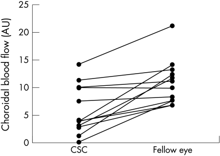
Methods: A complete clinical ophthalmological examination was performed using simultaneous fluorescein and indocyanine green (ICG) angiography with a confocal scanning laser ophthalmoscopy and the digital images analysed in 36 consecutive patients with acute CSC. To quantify the choroidal circulation, the foveal choroidal blood flow was measured in 11 patients using laser Doppler flowmetry.
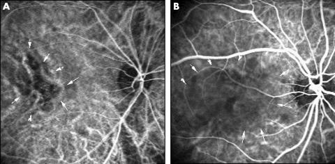
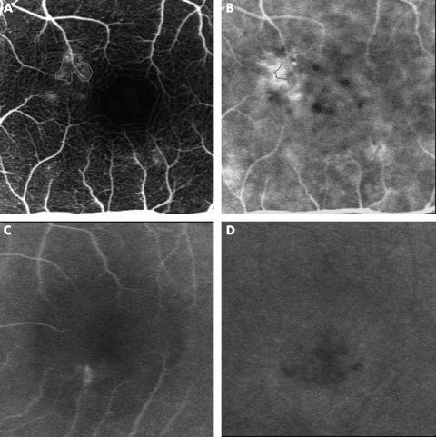
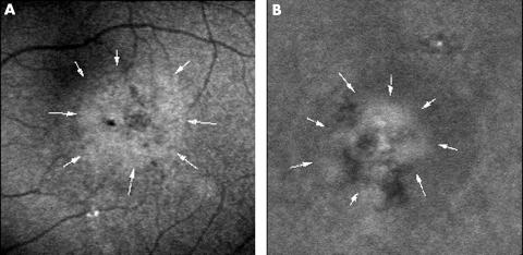
Results: Fluorescein angiography showed focal leakage from the retinal pigment epithelium in all patients. ICG angiography revealed delays in arterial filling in 27 eyes (75%), and fluorescein angiography showed small hypofluorescent points around the leakage in 27 eyes (75%). Abnormal choroidal hyperfluorescence was observed in 30 eyes (83%). The choroidal blood flow in eyes with CSC was 45% lower than in fellow eyes (p<0.01).
Conclusion: Decreased choroidal blood flow in CSC was demonstrated for the first time. The decreased choroidal blood flow might be correlated with the small, localised hypofluorescent areas, which may indicate non-perfused areas of the choriocapillaris that are frequently seen during ICG angiography.
Figures
References
-
- Gass JDM. Pathogenesis of disciform detachment of the neuroepithelium, II: idiopathic central serous chorioretinopathy. Am J Ophthalmol 1967;63:587–615. - PubMed
-
- Yannuzzi LA, Gitter KA, Schats H. Central serous chorioretinopathy. In: Yannuzzui LA, Gitter KA, Schats H, eds. The macula: a comprehensive text and atlas. Baltimore: Williams & Wilkins, 1979.
-
- Yannuzzi LA, Shakin JL, Fisher YL, et al. Peripheral retinal detachments and retinal pigment epithelial atrophic tracts secondary to central serous pigment epitheliopathy. Ophthalmology 1984;91:1554–72. - PubMed
-
- Spitznas M. Pathogenesis of central serous retinopathy: a new working hypothesis. Graefes Arch Clin Exp Ophthalmol 1986;224:321–4. - PubMed
-
- Marmor MF. New hypothesis on the pathogenesis and treatment of serous retinal detachment. Graefes Arch Clin Exp Ophthalmol 1988;226:548–52. - PubMed
MeSH terms
Substances
LinkOut - more resources
Full Text Sources
Medical