Extent of foveal tritanopia in diabetes mellitus
- PMID: 12770973
- PMCID: PMC1771723
- DOI: 10.1136/bjo.87.6.742
Extent of foveal tritanopia in diabetes mellitus
Abstract
Aim: To use a colour matching technique to test the hypothesis that the foveal tritanopic zone is increased in size in diabetes mellitus.
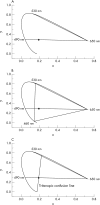
Method: A Wright tristimulus colorimeter was adapted for small field colour matching and colour matches were performed on bipartite fields in the range 12' to 60' of arc. The reference stimulus was 490 nm desaturated with 650 nm and the matching stimulus consisted of either two wavelengths (530 nm and 650 nm) or three (460 nm, 530 nm, and 650 nm). The size of the zone of foveal tritanopia was measured using two alternative forced choice presentations of dichromatic and trichromatic matches made by the observer for different field sizes. 21 diabetic and 12 controls performed the experiment.
Results: The results for the controls show a normal distribution, with a median foveal tritanopic zone of 18' of arc. The median for the diabetic patients was also 18' of arc, but the distribution showed a significant skew to the right. A non-parametric test shows a significant difference in comparison with the controls (p = 0.01), with several subjects having extensive zones of foveal tritanopia, reaching up to 1 degree.
Conclusions: In the majority of diabetic subjects the extent of foveal tritanopia is normal; however, there is good evidence that in a small number of subjects the size of the zone is significantly increased. This indicates S-cone pathway damage that is sufficiently severe to lead to dichromatic colour vision in the fovea.
Figures


Similar articles
-
Classical tritanopia.J Physiol. 1983 Feb;335:655-81. doi: 10.1113/jphysiol.1983.sp014557. J Physiol. 1983. PMID: 6603508 Free PMC article.
-
Study of foveal tritanopia.Percept Mot Skills. 1978 Jun;46(3 Pt 2):1319-27. doi: 10.2466/pms.1978.46.3c.1319. Percept Mot Skills. 1978. PMID: 308216
-
Silent substitution S-cone electroretinogram in subjects with diabetes mellitus.Ophthalmic Physiol Opt. 2005 Sep;25(5):392-9. doi: 10.1111/j.1475-1313.2005.00299.x. Ophthalmic Physiol Opt. 2005. PMID: 16101944
-
[Diabetes and color vision disorder detected by the Farnsworth 100 Hue test. Diabetic dyschromatopsia].J Fr Ophtalmol. 1990;13(10):506-10. J Fr Ophtalmol. 1990. PMID: 2081841 Review. French.
-
FURTHER STUDIES OF EXTRA-FOVEAL COLOUR METRICS.Opt Acta (Lond). 1963 Jul;10:257-84. doi: 10.1080/713817799. Opt Acta (Lond). 1963. PMID: 14073071 Review. No abstract available.
Cited by
-
Chromatic-achromatic perimetry in four clinic cases: Glaucoma and diabetes.Indian J Ophthalmol. 2015 Feb;63(2):146-51. doi: 10.4103/0301-4738.154392. Indian J Ophthalmol. 2015. PMID: 25827546 Free PMC article.
-
Long-Term Anatomic and Functional Outcomes after Macular Hole Surgery.J Ophthalmol. 2018 Nov 26;2018:3082194. doi: 10.1155/2018/3082194. eCollection 2018. J Ophthalmol. 2018. PMID: 30598845 Free PMC article.
References
-
- Kolb H, Lipetz L. The anatomical basis for colour vision in the vertebrate retina. In: Gouras P, ed. Vision and visual dysfunction. London: Macmillan, 1991:128–45.
-
- Konig A. Uber den menschlichen Sehpurpur und seine Bedeutung fur das Sehen. SB Akad Wiss Berlin 1894:577–98.
-
- Willmer E. Colour of small objects. Nature 1944;153:774–5.
-
- Willmer E, Wright W. Colour sensitivity of the fovea centralis. Nature 1945;156:119–21.
-
- Hartridge H. Colour vision of the fovea centralis. Nature 1945;155:391–2.
Publication types
MeSH terms
Grants and funding
LinkOut - more resources
Full Text Sources
Medical
