. 2003 May 26;161(4):769-77.
doi: 10.1083/jcb.200211094.

Dishevelled activates Ca2+ flux, PKC, and CamKII in vertebrate embryos

Affiliations

Dishevelled activates Ca2+ flux, PKC, and CamKII in vertebrate embryos

Laird C Sheldahl et al. J Cell Biol. .

Abstract

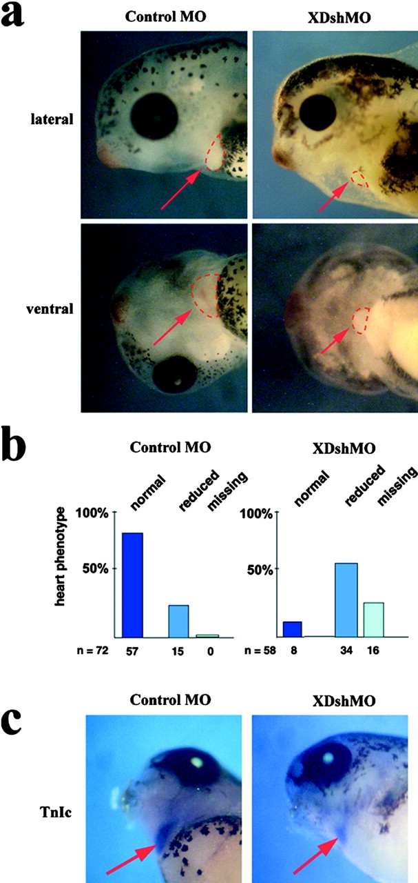
Wnt ligands and Frizzled (Fz) receptors have been shown to activate multiple intracellular signaling pathways. Activation of the Wnt-beta-catenin pathway has been described in greatest detail, but it has been reported that Wnts and Fzs also activate vertebrate planar cell polarity (PCP) and Wnt-Ca2+ pathways. Although the intracellular protein Dishevelled (Dsh) plays a dual role in both the Wnt-beta-catenin and the PCP pathways, its potential involvement in the Wnt-Ca2+ pathway has not been investigated. Here we show that a Dsh deletion construct, XDshDeltaDIX, which is sufficient for activation of the PCP pathway, is also sufficient for activation of three effectors of the Wnt-Ca2+ pathway: Ca2+ flux, PKC, and calcium/calmodulin-dependent protein kinase II (CamKII). Furthermore, we find that interfering with endogenous Dsh function reduces the activation of PKC by Xfz7 and interferes with normal heart development. These data suggest that the Wnt-Ca2+ pathway utilizes Dsh, thereby implicating Dsh as a component of all reported Fz signaling pathways.


Figures

Figure 1.
Figure 1.
XDshΔDIX activates PKC in a PTX- insensitive manner. Two-cell-stage Xenopus embryos were injected with RNAs encoding XPKCα–myc (0.5–1 ng) plus XDsh–GFP (0.5–1 ng), XDshΔDEP–GFP (0.5–1 ng), XDshΔDIX–GFP (0.5–1 ng), Rfz2 (0.5–1 ng), PTX (1–2 ng), and/or LacZ (as a control for normalizing levels of injected RNA, 0–4 ng) and cultured to stage 8, and animal caps were explanted. (a) XPKC (red) is localized primarily in the cytoplasm under control conditions. (b) Ectopic XDsh–GFP (green), which is localized to punctate structures, is relatively weak at activating translocation of XPKC (red) to the membrane. (c) XDshΔDEP–GFP (green) as well as XDshΔPDZ–GFP (not depicted) do not activate XPKC (red) membrane translocation. (c, inset) XDsh–GFP (lane 1) and XDshΔDIX–GFP (lane 2) are expressed equally, as detected by an anti-GFP Western blot. Although the protein in lane 2 migrates more rapidly on gels compared with lane 1 (not depicted), it is aligned with lane 1 to facilitate the comparison of signal intensities. (d) Expression of XDshΔDIX–GFP (green) activates translocation to the membrane of PKC (red). (e) Rfz2 (untagged) activates membrane translocation of XPKC (red). (f) Rfz-2–mediated membrane translocation of XPKC (red) is partially blocked by PTX. (g) The ability of XDshΔDIX–GFP (green) to induce membrane translocation of XPKC (red) is not blocked by PTX. (h) A schematic representing the three domains of Dsh discussed in the text.
Figure 2.
Figure 2.
XDshΔDIX activates Ca 2+ flux in a PTX-insensitive manner. Zebrafish embryos were injected with a Ca2+-sensitive dye along with respective RNAs and subjected to image analysis. In all panels, the image corresponds to the outline of a zebrafish embryo, depicting the spatial distribution of Ca2+ flux or lineage tracers as noted. (a) Control Ca2+ flux and (b) Rfz2 RNA–induced Ca2+ flux are low and high reference points, respectively (purple denotes low Ca2+ flux, and blue, green, yellow, and red denote increasing Ca2+ flux, respectively, as shown in the bar below b). (c) Unilateral injection of wild-type XDsh RNA produces a mild increase in Ca2+ transients compared with the control, a. (d) Lineage tracer coinjected with wild-type XDsh RNA establishes the spatial distribution of RNA after injection. (e) Injection of XDshΔDIX RNA produces a significant increase in Ca2+ transients in the region of f, the lineage tracer for XDshΔDIX RNA injection. (g) Ca2+ transients (blue) in embryos uniformly injected with Rfz2 RNA are reduced in regions expressing the A protomer of PTX comixed with lineage tracer, as seen in h. (i) Ca2+ transients in embryos uniformly injected with XWnt-5A RNA are similarly reduced in PTX-injected regions; PTX and tracer distribution shown in j. (k) Unilateral injection of XDshΔDIX RNA produces a significant increase in Ca2+ transients not blocked by PTX, with the spatial distribution of PTX shown in l.
Figure 3.
Figure 3.
XDshΔDIX activates CamKII activity in a PTX-insensitive manner. Two-cell-stage Xenopus embryos were injected with RNAs encoding Rfz2, XDsh–GFP, XDshΔDIX–GFP, or PTX and processed for an in vitro CamKII activity assay. Injection of wt XDsh–GFP RNA produces a mild increase in CamKII activity compared with control embryos, whereas Rfz2 and XDshΔDIX–GFP RNAs produce a twofold activation compared with control embryos. Activation of CamKII by XDshΔDIX–GFP, unlike Rfz2, was not sensitive to coexpression of PTX. CamKII is rarely induced above two to threefold in any system (for review see Kühl et al., 2000a).
Figure 4.
Figure 4.
Expression of the DEP domain of XDsh interferes with Xfz7-mediated activation of PKC. Two-cell-stage Xenopus embryos were injected with RNAs encoding XPKCα–myc (0.5–1 ng) and Xfz7 (0.5–1 ng) plus XDsh–GFP (0.5–1 ng), XDshΔDEP–GFP (0.5–1 ng), XDshΔDIX–GFP (0.5–1 ng), XDshΔPDZ–GFP (0.5–1 ng), XDEP–GFP (0.5–1 ng), and/or LacZ (as a control for normalizing levels of injected RNA, 0–4 ng) and cultured to stage 8, and animal caps were explanted. (a) XPKC (red) is localized to the plasma membrane when coexpressed with untagged Xfz7. (b) XDshΔDEP–GFP (green) and (c) XDshΔPDZ–GFP (green) do not block Xfz7-mediated membrane translocation of XPKC (red). (d) Both wt XDsh–GFP (green) and XPKC (red, yielding some yellow signal in areas of overlap with XDsh) are recruited to the plasma membrane upon coexpression of untagged Xfz7. (e) Expression of XDEP–GFP reduces membrane relocalization of XPKC by untagged Xfz7 (note increased red cytoplasmic XPKC signal compared with panel a), whereas XDEP–GFP itself is recruited to the plasma membrane by Xfz7 (green signal, appearing yellow at the membrane due to overlap with the red signal). (f) In the absence of ectopic Xfz7, XPKC (red) is localized throughout the cytoplasm, and XDEP–GFP (green) is localized diffusely throughout the cytoplasm (yellow denotes areas of overlapping signal).
Figure 5.
Figure 5.
XDshMO blocks activation of PKC by Xfz7. Fertilized Xenopus eggs were injected with XDshMO (4–8 ng) or control MO (4–8 ng MoZNLK), plus RNAs encoding Xfz7 (0.5–1 ng), XDsh–HA (0.5–1 ng), XPKCα–myc (shown in panels a–d, 0.5–1 ng), and/or GFP (as a control to normalize RNA levels, 0–2 ng) and cultured to stage 8, and animal caps were explanted. (a) Xfz7 activates membrane translocation of XPKC (white signal). (b) Injection of XDshMO inhibits Xfz7-mediated membrane translocation of XPKC (note increased cytoplasmic signal compared with panel a). (c) A control MO does not inhibit membrane translocation of XPKC by Xfz7. (d) The effects of XDshMO can be rescued by injecting XDsh RNA that does not contain the 5′ UTR targeted by the MO (compare increased membrane staining of XPKC with the XPKC signal in panel b). (e) Western blots from animal cap lysates show that XDshMO reduces XDsh levels below that of GFP- or control MO-injected caps when probed with an anti-Dsh antibody (anti– Dvl-1). Coinjection of XDsh–HA RNA and XDshMO brings XDsh levels back to those of control animal caps, whereas expression of a control protein, spectrin, is not altered by any injection. (f) Lysates from animal caps were processed for an in vitro PKC activity assay, and a representative experiment is shown. Injection of XDshMO reduces XPKC activity below that of control MO-injected caps, and this activity is rescued by coinjection of XDsh–HA RNA.
Figure 6.
Figure 6.
Dsh is required for Xenopu s heart development: effect of XDshMO on cardiac development. (a) Vegetal blastomeres of Xenopus eight-cell embryos were injected with a total of 50 ng of XDshMO or control MO, and the development of the heart was analyzed at stage 41–43 for a contractile phenotype. Lateral and ventral views of representative injected embryos are given. The contracting areas are indicated by the dashed red line and by a red arrow. (b) Summary of phenotypes of injected embryos. n represents the number of injected embryos and, below the bars, the number of embryos with the indicated heart phenotype. (c) Whole mount in situ hybridization against TnIc reveals weaker staining in XDshMO-injected embryos in comparison with embryos injected with the control MO.

References

    1. Adler, P.N., and H. Lee. 2001. Frizzled signaling and cell-cell interactions in planar polarity. Curr. Opin. Cell Biol. 13:635–640. - PubMed
    1. Axelrod, J.D., J.R. Miller, J.M. Shulman, R.T. Moon, and N. Perrimon. 1998. Differential recruitment of Dishevelled provides signaling specificity in the planar cell polarity and Wingless signaling pathways. Genes Dev. 12:2610–2622. - PMC - PubMed
    1. Boutros, M., and M. Mlodzik. 1999. Dishevelled: at the crossroads of divergent intracellular signaling pathways. Mech. Dev. 83:27–37. - PubMed
    1. Boutros, M., N. Paricio, D.I. Strutt, and M. Mlodzik. 1998. Dishevelled activates JNK and discriminates between JNK pathways in planar polarity and wingless signaling. Cell. 94:109–118. - PubMed
    1. Brown, J.D., S.E. Hallagan, L.L. McGrew, J.R. Miller, and R.T. Moon. 2000. The maternal Xenopus β-catenin signaling pathway, activated by frizzled homologs, induces goosecoid in a cell non-autonomous manner. Dev. Growth Differ. 42:347–357. - PubMed

Publication types

MeSH terms