. 2003 Jun 2;197(11):1501-10.
doi: 10.1084/jem.20022031. Epub 2003 May 27.

The role of intramolecular epitope spreading in the pathogenesis of endemic pemphigus foliaceus (fogo selvagem)

Affiliations

The role of intramolecular epitope spreading in the pathogenesis of endemic pemphigus foliaceus (fogo selvagem)

Ning Li et al. J Exp Med. .

Abstract

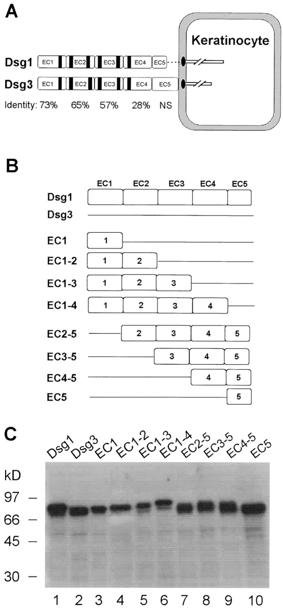
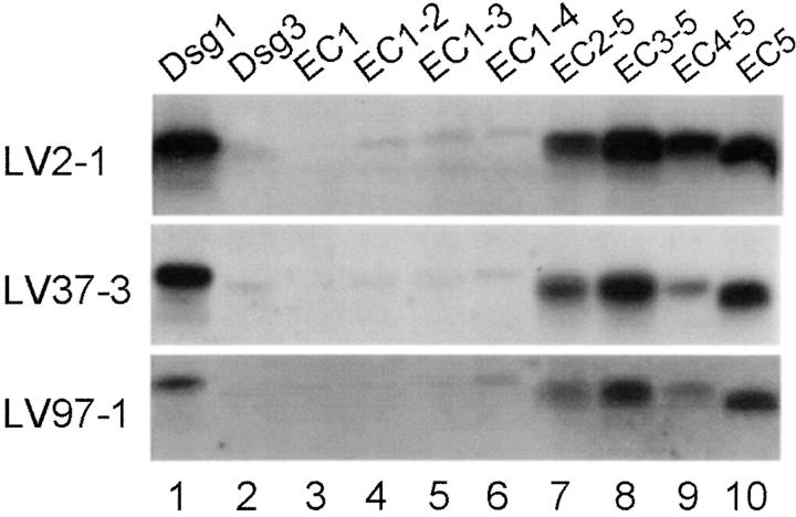
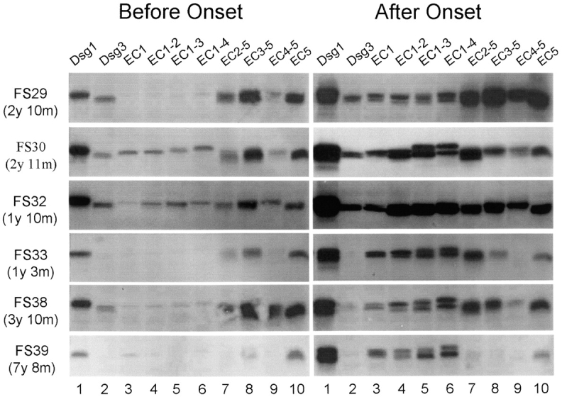
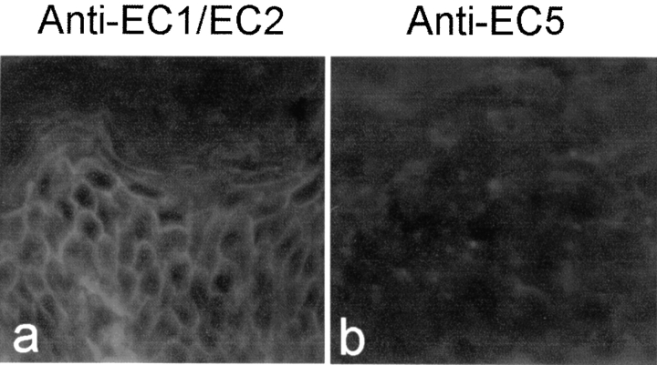
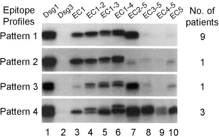
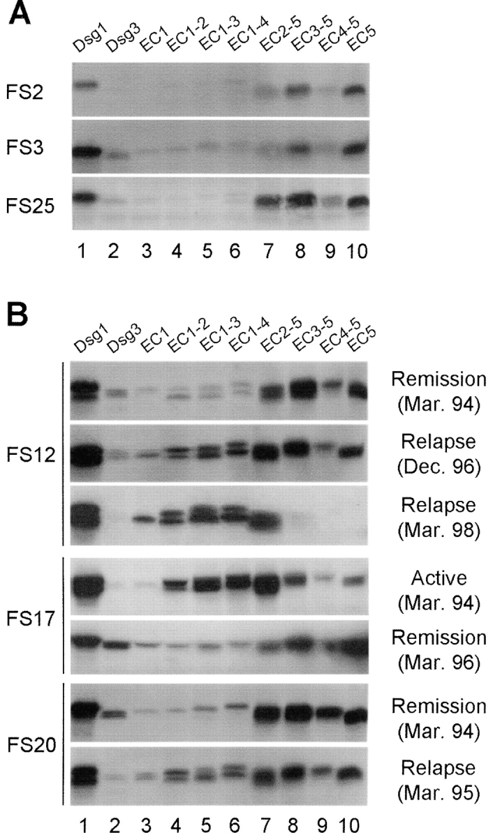
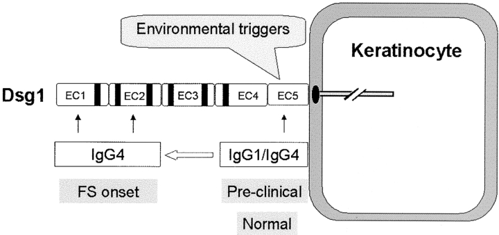
We report here a relationship between intramolecular epitope spreading and the clinical onset of the endemic form of pemphigus foliaceus in a Brazilian community with a high prevalence and incidence of the disease. Also known as Fogo Selvagem (FS), this disease is characterized by severe skin blistering and pathogenic anti-desmoglein-1 (Dsg1) autoantibodies. These autoantibodies bind the Dsg1 ectodomain and trigger keratinocyte cell detachment, the hallmark of FS. We show that (a) sera from FS patients in the preclinical stage recognized epitopes on the COOH-terminal EC5 domain of Dsg1, (b) disease onset was associated with the emergence of antibodies specific for epitopes on the NH2-terminal EC1 and EC2 domains, (c) all sera from FS patients with active disease recognized the EC1 and/or EC2 domains, and (d) sera from FS patients in remission showed reactivity restricted to EC5. These results suggest that anti-Dsg1 autoantibodies in FS are initially raised against the COOH-terminal EC5 domain of Dsg1 in individuals without skin disease; in genetically predisposed subjects the autoimmune response may then undergo intramolecular epitope spreading toward epitopes on the NH2-terminal EC1 and EC2 domains of Dsg1 leading to disease onset. Moreover, intramolecular epitope spreading may also modulate remissions and relapses of FS.


Figures

Figure 1.
Figure 1.
Schematic diagram and baculovirus production of Dsg1, Dsg3, and chimeric antigens. (A) Molecular structure of Dsg1 and Dsg3. Dsg1 and Dsg3 are structurally similar desmosomal transmembrane glycoproteins. The extracellular domain of the two molecules contains four cadherin-like domains (EC1 to EC4) and a membrane-proximal domain (EC5). Sequence identities between the cadherin repeats of Dsg1 and Dsg3 are indicated. NS indicates that sequence homology between the two molecules in the EC5 domain is not significant (reference 39). The black stripes represent six putative Ca2+ binding motifs found in the cadherin repeats. (B) Diagram of the recombinant Dsg1, Dsg3, and eight chimeric proteins. We constructed the full-length extracellular domains of Dsg1, Dsg3, and eight hybrid molecules that contained various combinations of Dsg1 extracellular domains (open box) with the corresponding Dsg3 (straight line) backbone. All recombinant proteins contain the endogenous signal sequence, a propeptide, and the extracellular domain of Dsg1/Dsg3 fused with a COOH-terminal His tag. (C) Production of the 10 recombinant proteins in the baculovirus expression system. The culture supernatants containing various secreted recombinant proteins (labeled on top of the figure) were collected and subjected to immunoblotting using HRP-labeled monoclonal antibody against the common His tag. Molecular weight standards are shown at left.
Figure 2.
Figure 2.
Analysis of anti-Dsg1 antibodies from normal subjects living in an area endemic for FS. Each serum sample was immunoprecipitated with 10 recombinant proteins (labeled on top of each panel), respectively, and subjected to immunoblotting using a monoclonal antibody against the His tag. IP results from three normal individuals living in the Limao Verde of Brazil are shown.
Figure 3.
Figure 3.
Antibody specificities from FS patients before and after the clinical onset of disease. Sera from six patients before (left panel) or after (right panel) clinical disease onset were analyzed by IP. Left panel: the time intervals between the preclinical sera and disease onset are indicated at left. y: year(s); m: months. Right panel: sera from patients FS29, FS32, and FS33 were collected after 3, 2, or 9 mo, respectively after the first symptoms of FS observed; sera from patients FS30, FS38, and FS39 were collected within the same month of disease onset.
Figure 4.
Figure 4.
Indirect IF analysis of anti-EC1/EC2 and anti-EC5 sera on the human epidermis. Left panel: a serum from a FS patient with anti-Dsg1 antibodies specific for the EC1 and EC2 domains. Right panel: a normal serum with anti-Dsg1 antibodies specific for the EC5 domain of Dsg1.
Figure 5.
Figure 5.
Epitope profiles of anti-Dsg1 antibodies from patients with active disease. IP results from four representative FS patients with active disease are shown. The number of sera within this group (n = 14) showing similar reaction patterns is indicated at right.
Figure 6.
Figure 6.
Antibody specificities from FS patients during the course of disease. (A) IP results from three FS patients in long-term clinical remission. (B) IP results from sequential sera samples obtained from three representative patients (FS12, FS17, and FS20) during the course of disease. Clinical outcomes and bleeding dates are labeled at right.
Figure 7.
Figure 7.
A model of the immunopathogenic pathway of FS. Environmental factor(s) trigger the production of nonpathogenic IgG1 and IgG4 antibodies against the EC5 domain of Dsg1. In certain genetically predisposed individuals, intramolecular epitope spreading results in the generation of predominantly IgG4 antibodies, against the EC1 and EC2 domains of Dsg1, that induce disease.

References

    1. Rock, B., C.R. Martins, A.N. Theofilopoulos, R.S. Balderas, G.J. Anhalt, R.S. Labib, S. Futamura, E.A. Rivitti, and L.A. Diaz. 1989. The pathogenic effect of IgG4 autoantibodies in endemic pemphigus foliaceus (fogo selvagem). N. Engl. J. Med. 320:1463–1469. - PubMed
    1. Seishima, M., Y. Iwasaki-Bessho, Y. Itoh, Y. Nozawa, M. Amagai, and Y. Kitajima. 1999. Phosphatidylcholine-specific phospholipase C, but not phospholipase D, is involved in pemphigus IgG-induced signal transduction. Arch. Dermatol. Res. 291:606–613. - PubMed
    1. Mahoney, MyG., Z. Wang, K. Rothenberger, P.J. Koch, M. Amagai, and J.R. Stanley. 1999. Explanations for the clinical and microscopic localization of lesions in pemphigus foliaceus and vulgaris. J. Clin. Invest. 103:461-468. - PMC - PubMed
    1. Kowalczyk, A.P., J.E. Anderson, J.E. Borgwardt, T. Hashimoto, J.R. Stanley, and K.J. Green. 1995. Pemphigus sera recognize conformationally sensitive epitopes in the amino-terminal region of desmoglein-1. J. Invest. Dermatol. 105:147–152. - PubMed
    1. Eyre, R.W., and J.R. Stanley. 1987. Human autoantibodies against a desmosomal protein complex with a calcium-sensitive epitope are characteristic of pemphigus foliaceus patients. J. Exp. Med. 165:1719–1724. - PMC - PubMed

Publication types

MeSH terms