Comparative Study
. 2003 Jun 1;31(11):2778-85.
doi: 10.1093/nar/gkg376.

Domain mapping of Escherichia coli RecQ defines the roles of conserved N- and C-terminal regions in the RecQ family

Affiliations
Comparative Study

Domain mapping of Escherichia coli RecQ defines the roles of conserved N- and C-terminal regions in the RecQ family

Douglas A Bernstein et al. Nucleic Acids Res. .

Abstract

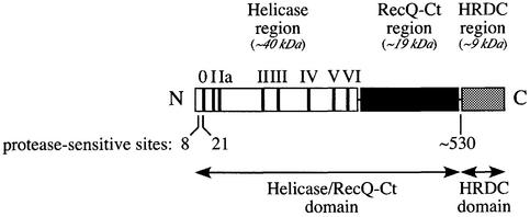
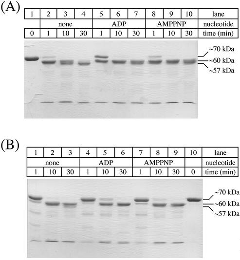
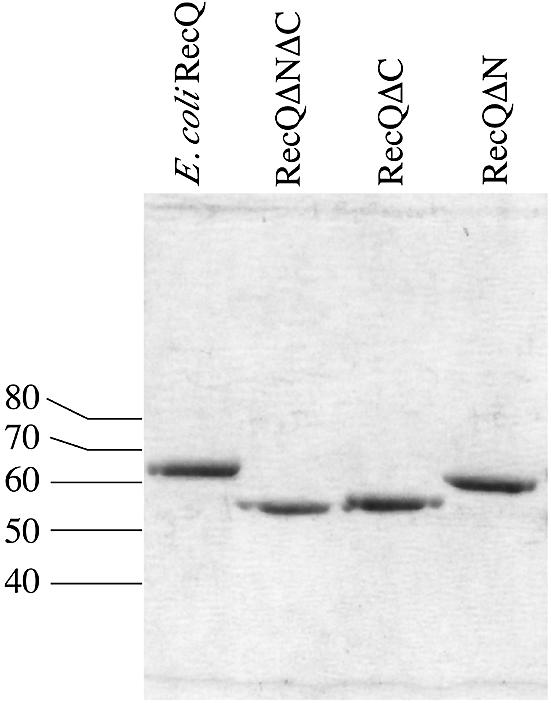
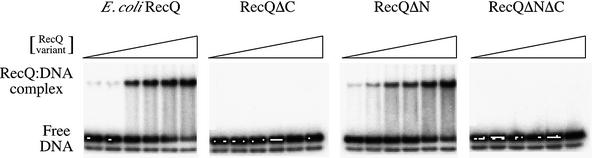
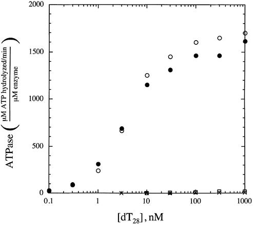
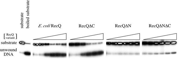
RecQ DNA helicases function in DNA replication, recombination and repair. Although the precise cellular roles played by this family of enzymes remain elusive, the importance of RecQ proteins is clear; mutations in any of three human RecQ genes lead to genomic instability and cancer. In this report, proteolysis is used to define a two-domain structure for Escherichia coli RecQ, revealing a large (approximately 59 kDa) N-terminal and a small (approximately 9 kDa) C-terminal domain. A short N-terminal segment (7 or 21 residues) is also shown to be sensitive to proteases. The effects of removing these regions of RecQ are tested in vitro. Removing 21 N-terminal residues from RecQ severely diminishes its DNA-dependent ATPase and helicase activities, but does not affect its ability to bind DNA in electrophoretic mobility shift assays. In contrast, removing the approximately 9 kDa C-terminal domain from RecQ results in a fragment with normal levels of ATPase and helicase activity, but that has lost the ability to stably associate with DNA. These results establish the biochemical roles of an N-terminal sequence motif in RecQ catalytic function and for the C-terminal RecQ domain in stable DNA binding.


Figures

Figure 1
Figure 1
Schematic diagram of E.coli RecQ. Conserved regions, Helicase (white), RecQ-Ct (black) and HRDC (gray) (20) are shown as boxes. The locations of the conserved helicase motifs (I–VI) and a proteolytically labile motif identified in this study (motif 0) are indicated with bars in the Helicase box. The approximate molecular weight of each region is listed. Protease-sensitive sites and structural domains identified in this study are indicated below the schematic.
Figure 2
Figure 2
Limited proteolysis of E.coli RecQ. Purified E.coli RecQ (14 µM) was treated with 0.14 µM chymotrypsin (A) or subtilisin (B) as described in the Materials and Methods, quenched at the indicated times, separated by SDS–PAGE, and stained with Coomassie Brilliant Blue. In some reactions, ADP or AMPPNP was included at 0.1 mM. Aliquots of E.coli RecQ prior to the addition of protease are shown (A, lane 1, and B, lane 10). Molecular weight estimates from SDS–PAGE analyses are indicated.
Figure 3
Figure 3
SDS–PAGE analysis of purified E.coli RecQ variants. Escherichia coli RecQ, RecQΔN, RecQΔC and RecQΔNΔC were purified as described in the Materials and Methods. Equimolar amounts of each were separated on a 10% SDS–PAGE gel and stained with Coomassie Brilliant Blue. Molecular weights from protein standards are indicated.
Figure 4
Figure 4
EMSA analysis of DNA binding by E.coli RecQ, RecQΔN, RecQΔC and RecQΔNΔC. Proteins at 8, 16, 31, 62, 125 or 250 nM were incubated with a ∼1 nM radiolabeled DNA substrate as described in the Materials and Methods and protein:DNA complexes were separated from free DNA by PAGE on a 6% non-denaturing gel. Radiolabeled DNA was observed using a phosphorimager. A small amount of the radiolabeled DNA strand from the ds substrate is observed in its ss, unannealed form in each lane. Free DNA and RecQ:DNA complex bands are indicated. Binding experiments were performed multiple times and the data shown are representative of all observations.
Figure 5
Figure 5
Analysis of DNA-dependent ATPase activity in E.coli RecQ (filled circles), RecQΔN (‘X’ symbols), RecQΔC (open circles) and RecQΔNΔC (open boxes). Proteins were incubated with indicated concentrations of dT28 in the presence of 1 mM ATP in a coupled ATP regeneration/NADH oxidation system (47) as described in the Materials and Methods. For full-length RecQ and RecQΔC, protein concentrations were 1–8 nM whereas the weakly active RecQΔN and RecQΔNΔC variants required 50–500 nM enzyme concentrations to produce a measurable ATPase rate. Data points are normalized to the specific enzyme concentration used in each measurement.
Figure 6
Figure 6
DNA helicase activity in E.coli RecQ, RecQΔN, RecQΔC and RecQΔNΔC. Proteins at 0.001, 0.01, 0.1, 1, 10 or 100 nM were incubated with a ∼1 nM radiolabeled DNA substrate in the presence of 1 mM ATP as described in the Materials and Methods and unwound DNA was separated from substrate by PAGE on a 12% non-denaturing gel. Radiolabeled DNA was observed using a phosphorimager. Substrate and boiled substrate controls are indicated. DNA unwinding experiments were performed multiple times and the data shown are representative of all observations.
Figure 7
Figure 7
Alignment of a conserved sequence motif (motif 0) in 12 bacterial RecQ proteins. The consensus ‘motif 0’ amino acid sequence is written with invariant (underlined) and conserved (italicized) residues shown using single-letter amino acid nomenclature, and with non-conserved residues given as ‘x’. Numbers in parentheses are the number of amino acids either N-terminal to motif 0 or between motif 0 and motif I. Motif 0 is shared among all predicted bacterial RecQ proteins with only a subset shown here to demonstrate the most diverse sequences observed.

Similar articles

Cited by

References

    1. Frei C. and Gasser,S.M. (2000) RecQ-like helicases: the DNA replication checkpoint connection. J. Cell Sci., 113 (Pt 15), 2641–2646. - PubMed
    1. Karow J.K., Wu,L. and Hickson,I.D. (2000) RecQ family helicases: roles in cancer and aging. Curr. Opin. Genet. Dev., 10, 32–38. - PubMed
    1. Nakayama H., Nakayama,K., Nakayama,R., Irino,N., Nakayama,Y. and Hanawalt,P.C. (1984) Isolation and genetic characterization of a thymineless death-resistant mutant of Escherichia coli K12: identification of a new mutation (recQ1) that blocks the RecF recombination pathway. Mol. Gen. Genet., 195, 474–480. - PubMed
    1. Kolodner R., Fishel,R.A. and Howard,M. (1985) Genetic recombination of bacterial plasmid DNA: effect of RecF pathway mutations on plasmid recombination in Escherichia coli. J. Bacteriol., 163, 1060–1066. - PMC - PubMed
    1. Luisi-DeLuca C., Lovett,S.T. and Kolodner,R.D. (1989) Genetic and physical analysis of plasmid recombination in recB recC sbcB and recB recC sbcA Escherichia coli K-12 mutants. Genetics, 122, 269–278. - PMC - PubMed

Publication types