. 2003 Jun 1;31(11):e64.
doi: 10.1093/nar/gng063.

In vitro analysis of nuclear mRNA export using molecular beacons for target detection

Affiliations

In vitro analysis of nuclear mRNA export using molecular beacons for target detection

Ralph H Kehlenbach. Nucleic Acids Res. .

Abstract

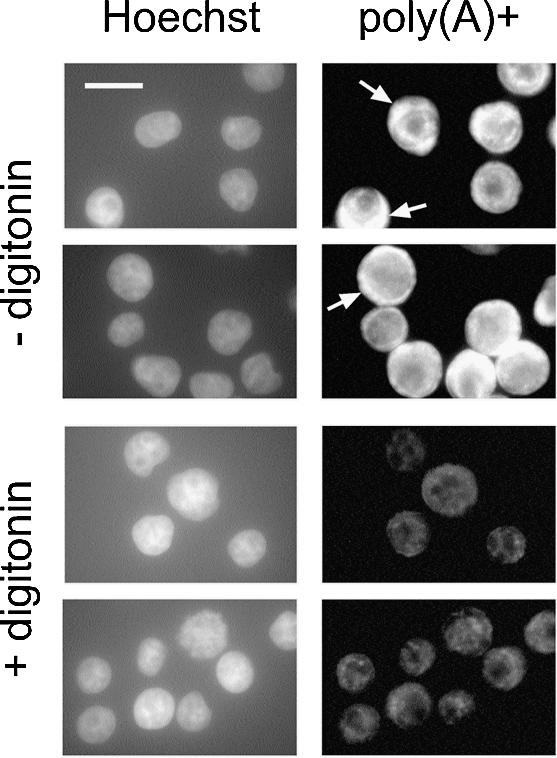
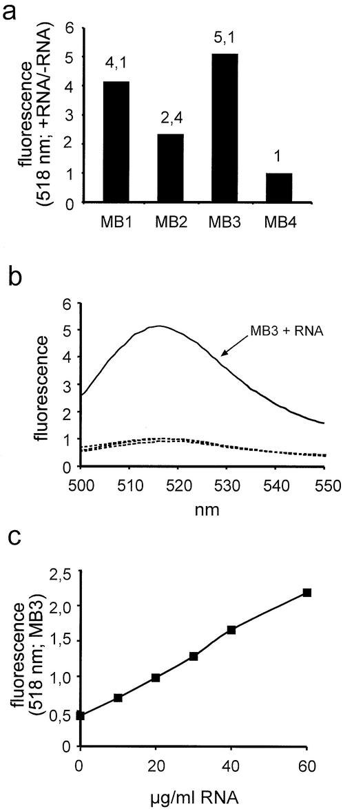
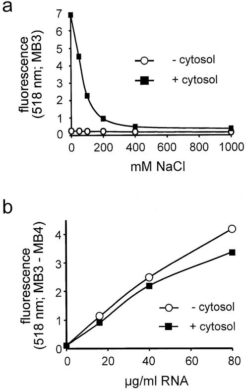
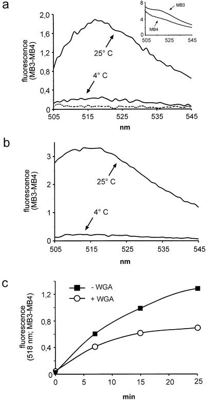
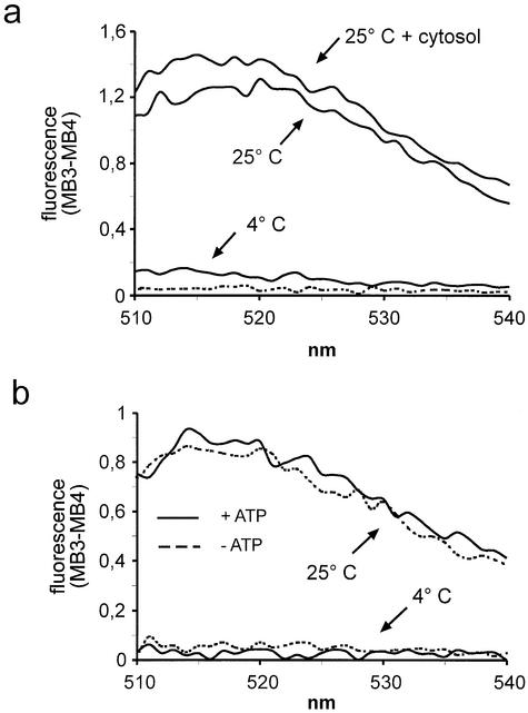
A detailed molecular characterization of nuclear mRNA export will require an in vitro system, allowing a biochemical reconstitution of transport. To this end, an mRNA export assay has been developed using digitonin-permeabilized HeLa cells and 2'-O-methyl oligoribonucleotide molecular beacons for target detection. These probes allow the homogeneous detection of poly(A)+ RNA at subnanomolar concentrations in the presence of cytosol, without the need for RNA purification and time-consuming methods like northern blotting or RT-PCR. Nuclear export of endogenous mRNA in permeabilized cells occurs in a time- and temperature-dependent manner and can be inhibited by wheat germ agglutinin, indicative of specific transport through nuclear pore complexes. Nuclear export in vitro is insensitive to the depletion of ATP and does not depend on the addition of cytosolic factors, suggesting that shuttling proteins are not required for efficient transport. This is the first demonstration of molecular beacons as powerful tools for the analysis of nucleocytoplasmic RNA transport.


Figures

Figure 1
Figure 1
Selective release of poly(A)+ RNA from the cytoplasm by digitonin permeabilization. In situ hybridizations were performed on intact cells (– digitonin) or permeabilized cells (+ digitonin). Nuclei were visualized by Hoechst staining (left). Some cells with a strong cytoplasmic staining for poly(A)+ RNA (– digitonin; right) are marked by arrows. Bar 20 µm.
Figure 2
Figure 2
Detection of mRNA with the help of molecular beacons. (a) Molecular beacons MB1–MB4 were incubated with or without 100 µg of purified total RNA, the resulting fluorescence was recorded at 518 nm, and plotted as the ratio of the data obtained under the two conditions (+ RNA/– RNA). (b) Molecular beacons MB3 or MB4 were incubated with or without 100 µg of purified RNA and the fluorescence emission spectra were recorded. Only MB3 in the presence of RNA (solid line) exhibited a signal above background. The three other spectra were basically identical (three dotted lines: MB3 – RNA, MB4 ± RNA). (c) Molecular beacon MB3 was incubated with increasing concentrations of purified total RNA and the resulting fluorescence was recorded at 518 nm.
Figure 3
Figure 3
Detection of RNA in the presence of cytosolic proteins. (a) Molecular beacon MB3 was incubated in the presence or absence of cytosol (1 mg/ml) with increasing concentrations of NaCl and the resulting fluorescence at 518 nm was plotted. (b) Molecular beacons MB3 or MB4 were incubated with increasing concentrations of total RNA in the absence or presence of 0.5 mg/ml cytosol. The background signal (MB3) was subtracted from the specific signal (MB3) and the resulting fluorescence at 518 nm was normalized to the sample with no RNA in the absence of cytosol.
Figure 4
Figure 4
Nuclear mRNA export in vitro. (a) Reactions were performed in the presence of cytosol at 4 or 25°C, as indicated. Fluorescence emission spectra were plotted after background subtraction (MB3 – MB4). The inset shows the original curves, prior to subtraction. The dotted line represents a reaction at 25°C without cells, indicating that the cytosol alone did not contribute significantly to the signal. (b) As (a) but after partial purification of exported RNA by phenol–chloroform extraction. (c) Kinetic analysis of nuclear export in the absence or presence of WGA. Nuclear export reactions were performed with (circles) or without (squares) 200 µg/ml WGA for up to 25 min.
Figure 5
Figure 5
Nuclear export in vitro does not depend on exogenous cytosol or ATP. (a) Nuclear export reactions were performed at 4 or 25°C with or without cytosol (1 mg/ml), as indicated. The dotted line represents a reaction at 25°C with cytosol but without cells. (b) Reactions were performed in the presence of an ATP-regenerating system (solid lines) or an ATP-depleting system (dotted lines) at either 4 or 25°C, as indicated.

References

    1. Görlich D. and Kutay,U. (1999) Transport between the cell nucleus and the cytoplasm. Annu. Rev. Cell. Dev. Biol., 15, 607–660. - PubMed
    1. Macara I.G. (2001) Transport into and out of the nucleus. Microbiol. Mol. Biol. Rev., 65, 570–594. - PMC - PubMed
    1. Weis K. (2003) Regulating access to the genome: nucleocytoplasmic transport throughout the cell cycle. Cell, 112, 441–451. - PubMed
    1. Fahrenkrog B., Stoffler,D. and Aebi,U. (2001) Nuclear pore complex architecture and functional dynamics. Curr. Top. Microbiol. Immunol., 259, 95–117. - PubMed
    1. Kalab P., Weis,K. and Heald,R. (2002) Visualization of a Ran-GTP gradient in interphase and mitotic Xenopus egg extracts. Science, 295, 2452–2456. - PubMed

Publication types