. 2003 Jun 2;88(11):1800-7.
doi: 10.1038/sj.bjc.6600984.

Apoptosis induction in renal cell carcinoma by TRAIL and gamma-radiation is impaired by deficient caspase-9 cleavage

Affiliations

Apoptosis induction in renal cell carcinoma by TRAIL and gamma-radiation is impaired by deficient caspase-9 cleavage

U Ramp et al. Br J Cancer. .

Abstract

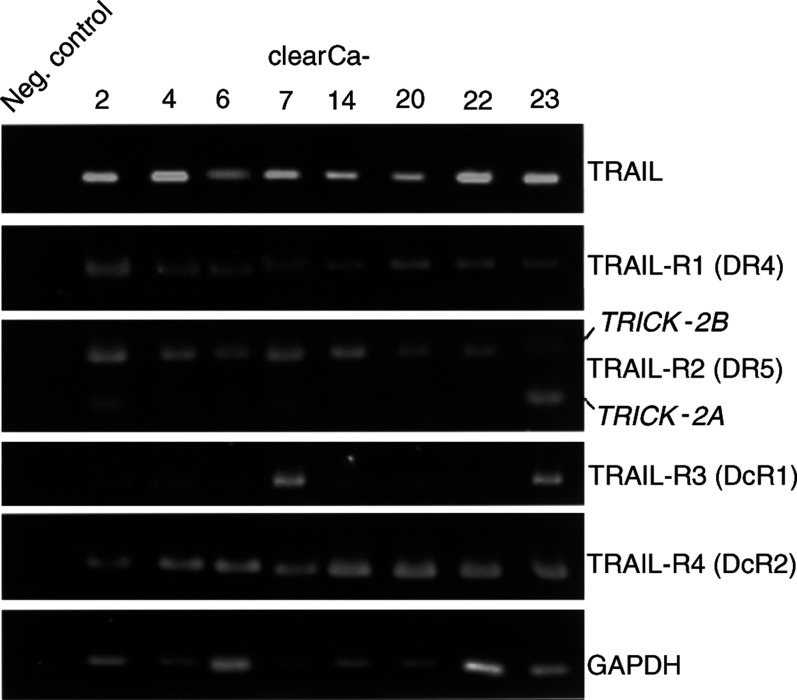
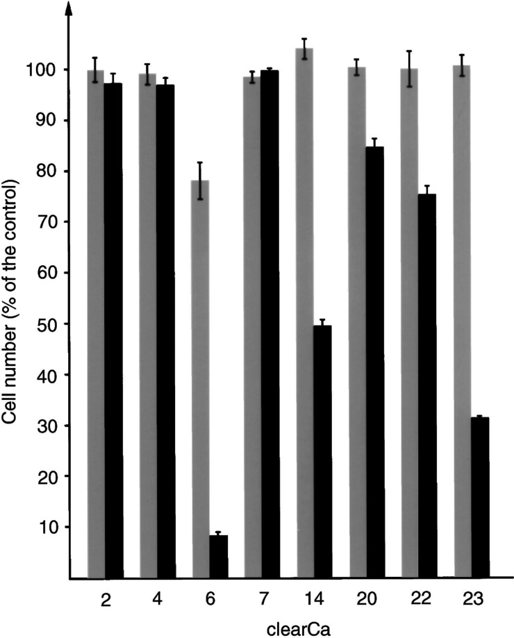
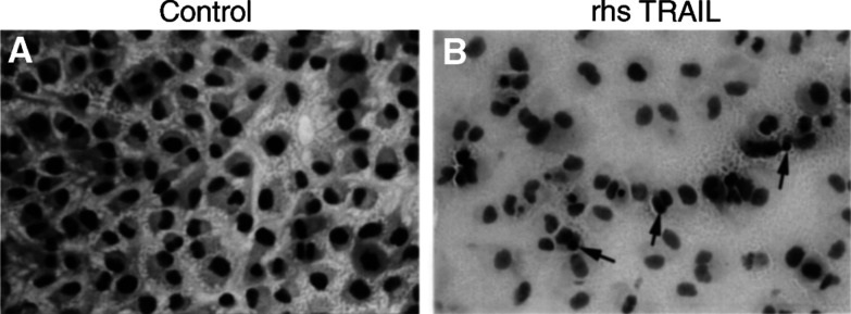
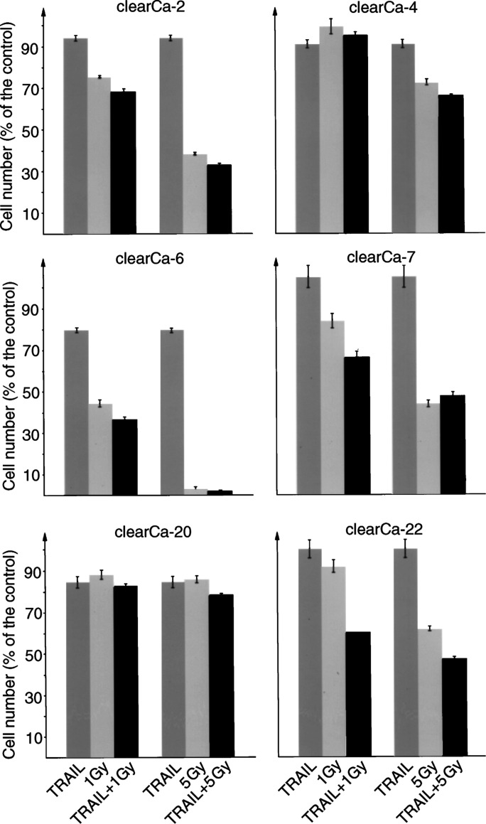
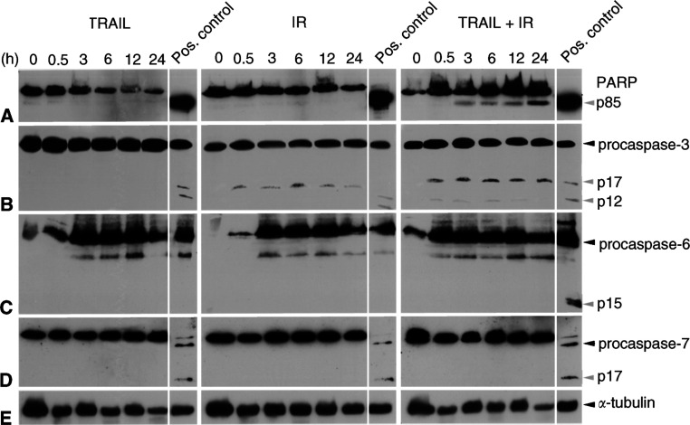
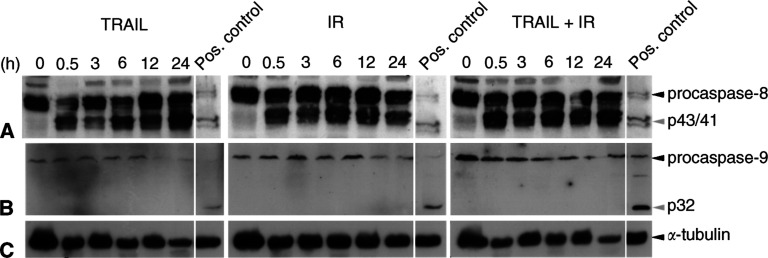
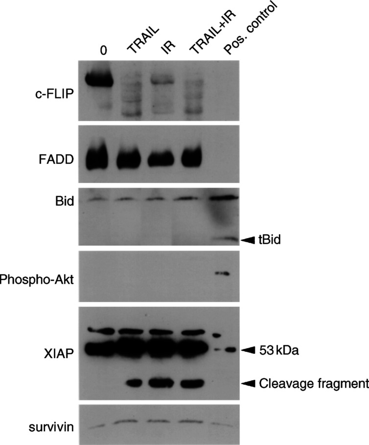
TNF-related apoptosis-inducing ligand (TRAIL APO-2L) is a member of the TNF family and induces apoptosis in cancer cells without affecting most non-neoplastic cells. The present investigation is focused on apoptosis induction by combined exposure to TRAIL and ionising radiation (IR) in human renal cell carcinoma (RCC) cell lines. Here, we demonstrate that all RCC cell lines coexpress TRAIL and the death-inducing receptors, TRAIL-R1 and TRAIL-R2. Exposure to TRAIL alone induced marked apoptosis in three out of eight RCC cell lines. Combined exposure to TRAIL and IR resulted in a sensitisation to TRAIL-induced apoptosis in one RCC cell line only. Enhanced apoptosis induction by TRAIL in combination with IR was paralleled by an increase in PARP cleavage and activation of executioner caspase-3, whereas caspases-6 and -7 were not involved. Moreover, exposure to TRAIL and/or IR resulted in a marked activation of initiator caspase-8, possibly augmented by the observed reduction of inhibitory c-FLIP expression. In contrast to other tumour types, activation of initiator caspase-9 was not detectable in our RCC model system after exposure to TRAIL and/or IR. This lack of caspase-9 activation might be related to an impaired 'crosstalk' with the caspase-8 pathway as suggested by the missing Bid cleavage and to the appearance of an XIAP cleavage product known to inhibit caspase-9 activation. Deficient activation of caspase-9, therefore, might contribute to the clinically known resistance of human RCC against IR and also argues against an effective combination therapy with TRAIL and IR in this tumour type.


Figures

Figure 1
Figure 1
Expression of TRAIL and its receptors in eight human RCC cell lines by RT–PCR. TRICK-2A and TRICK-2B are alternative splicing variants of TRAIL-R2. GAPDH detection was used as control for integrity of RNA and RT–PCR reactions.
Figure 2
Figure 2
Response of human RCC cell lines to TRAIL-induced cell death. Cells were cultured for 72 h in the presence of TRAIL (grey bars:10 ng ml−1 TRAIL; black bars: 100 ng ml−1 TRAIL) and the number of surviving, nonapoptotic cells is demonstrated in percentage of viable cells in the control (bars represent the s.d).
Figure 3
Figure 3
Marked induction of apoptosis (arrows) and marked reduction of cell density in clearCa-6 cells after exposure to TRAIL (100 ng ml−1) (B) when compared to the control (A). (Before TRAIL treatment, about 2% of the cells exhibited morphological features of apoptosis. The proportion of apoptotic cells after TRAIL treatment, however, cannot exactly be determined, because most apoptotic cells detached from the bottom of the culture flask and formed a not exactly quantifiable mass of cell detritus in the supernatant).
Figure4
Figure4
Sensitisation to TRAIL-induced apoptosis by IR in one (clearCa-22) out of six human RCC cell lines. RCC cell lines were cultured for 72 h in the presence of TRAIL (10 ng ml−1) or IR (1 or 5 Gy) alone or in combination and the number of surviving, nonapoptotic cells is demonstrated in percentage of viable cells in the control (bars represent the s.d).
Figure 5
Figure 5
Western blot analysis demonstrating marked cleavage of PARP (p85; A) and procaspase-3 (p17/p12; B) into its active fragments after combined exposure of clearCa-22 cells to TRAIL (10 ng ml−1) and IR (1 Gy). Exposure to TRAIL or IR alone did not result in increased cleavage. No cleavage products of procaspases-6 (p15; C) and -7 (p17; D) in clearCa-22 cells after exposure to TRAIL or IR alone and in combination. (cleavage of PARP as well as the procaspase-3, -6, and -7 in J16 cells after exposure to CH11 (500 ng ml−1) was used as a positive control). Expression of α-tubulin (E) shows equal amounts of protein loaded in each lane.
Figure 6
Figure 6
Western blot analysis demonstrating marked cleavage of procaspase-8 (A) into its active p43- and p41-fragments after exposure of clearCa-22 cells to TRAIL (10 ng ml−1) or IR (1 Gy) alone and in combination. No cleavage of procaspase-9 (B) in clearCa-22 cells after exposure to TRAIL or IR alone and in combination. (cleavage of procaspase-8 and -9 in J16 cells after exposure to CH11 (500 ng ml−1) was used as a positive control). Expression of α-tubulin (C) shows equal amounts of protein loaded in each lane.
Figure 7
Figure 7
Western blot analysis of apoptosis-regulating proteins in clearCa-22 after exposure to TRAIL (10 ng ml−1) or IR (1 Gy) alone and in combination for 12 h. Reduced expression of c-FLIP after exposure to TRAIL and/or IR, whereas expression of FADD and survivin remained unchanged when compared with the control (0). No cleavage of Bid into tBid and absence of active phosphorylated Akt protein after exposure to TRAIL and/or IR. High levels of XIAP protein (53 kDa) before and after exposure to TRAIL and/or IR and appearance of an XIAP cleavage product after exposure to TRAIL and/or IR. (Cleavage of Bid into tBid in J16 cells after exposure to CH11 was used as a positive control; expression of phosphorylated Akt in PDGF-induced NIH-3T3 cells was used as a positive control, as recommended by the manufacturer).

References

    1. Ashkenazi A, Pai RC, Fong S, Leung S, Lawrence DA, Marsters SA, Blackie C, Chang L, McMurtrey AE, Hebert A, DeForge L, Koumenis IL, Lewis D, Harris L, Bussiere J, Koeppen H, Shahrokh Z, Schwall RH (1999) Safety and antitumor activity of recombinant soluble Apo2 ligand. J Clin Invest 104: 155–162 - PMC - PubMed
    1. Belka C, Rudner J, Wesselborg S, Stepczynska A, Marini P, Lepple-Wienhues A, Faltin H, Bamberg M, Budach W, Schulze-Osthoff K (2000) Differential role of caspase-8 and BID activation during radiation- and CD95-induced apoptosis. Oncogene 19: 1181–1190 - PubMed
    1. Belka C, Schmid B, Marini P, Durand E, Rudner J, Faltin H, Bamberg M, Schulze-Osthoff K, Budach W (2001) Sensitization of resistant lymphoma cells to irradiation-induced apoptosis by the death ligand TRAIL. Oncogene 20: 2190–2196 - PubMed
    1. Brown JM, Wouters BG (1999) Apoptosis, p53, and tumor cell sensitivity to anticancer agents. Cancer Res 59: 1391–1399 - PubMed
    1. Chinnaiyan AM, Prasad U, Shankar S, Hamstra DA, Shanaiah M, Chenevert TL, Ross BD, Rehemtulla A (2000) Combined effect of tumor necrosis factor-related apoptosis-inducing ligand and ionizing radiation in breast cancer therapy. Proc Natl Acad Sci USA 97: 1754–1759 - PMC - PubMed

Publication types

MeSH terms

Substances