Comparative Study
. 2003 Jun 2;22(11):2571-82.
doi: 10.1093/emboj/cdg258.

Structural basis for SH3 domain-mediated high-affinity binding between Mona/Gads and SLP-76

Affiliations
Comparative Study

Structural basis for SH3 domain-mediated high-affinity binding between Mona/Gads and SLP-76

Maria Harkiolaki et al. EMBO J. .

Abstract

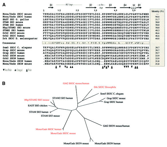
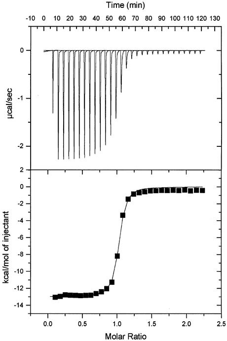
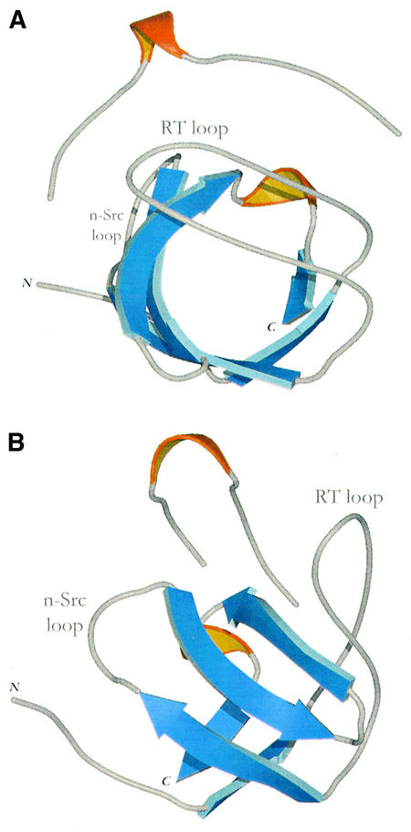
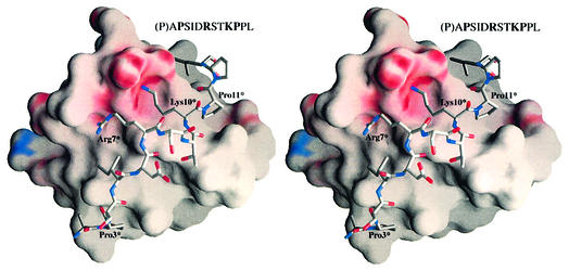
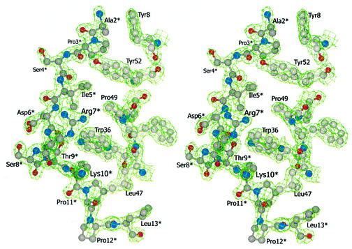
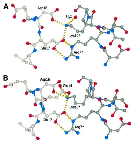
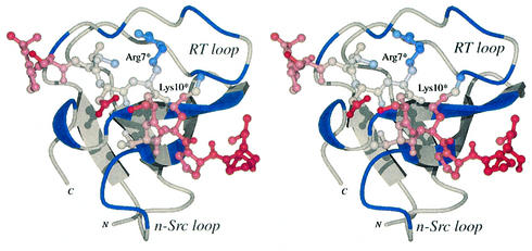
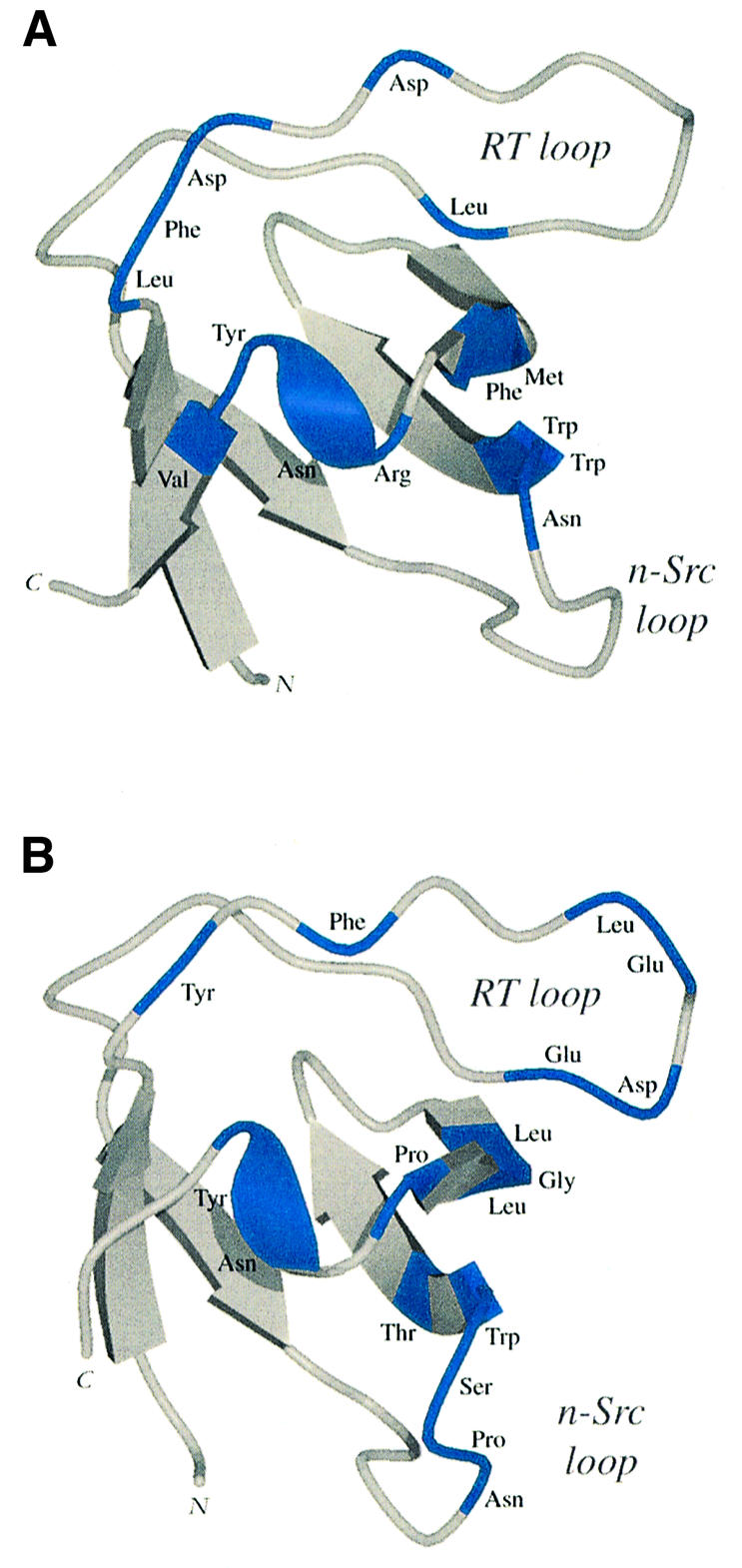
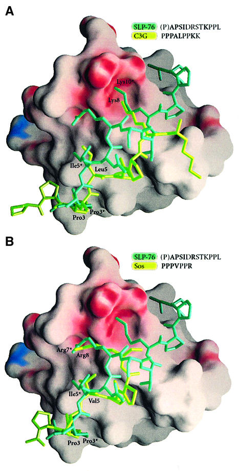
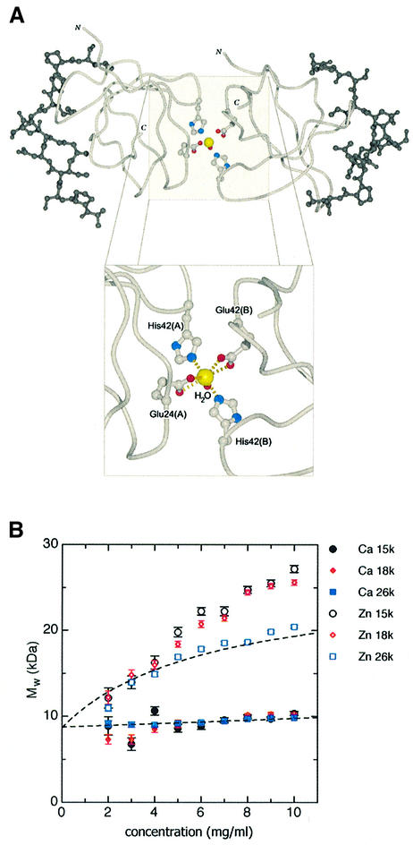
SH3 domains are protein recognition modules within many adaptors and enzymes. With more than 500 SH3 domains in the human genome, binding selectivity is a key issue in understanding the molecular basis of SH3 domain interactions. The Grb2-like adaptor protein Mona/Gads associates stably with the T-cell receptor signal transducer SLP-76. The crystal structure of a complex between the C-terminal SH3 domain (SH3C) of Mona/Gads and a SLP-76 peptide has now been solved to 1.7 A. The peptide lacks the canonical SH3 domain binding motif P-x-x-P and does not form a frequently observed poly-proline type II helix. Instead, it adopts a clamp-like shape around the circumfence of the SH3C beta-barrel. The central R-x-x-K motif of the peptide forms a 3(10) helix and inserts into a negatively charged double pocket on the SH3C while several other residues complement binding through hydrophobic interactions, creating a short linear SH3C binding epitope of uniquely high affinity. Interestingly, the SH3C displays ion-dependent dimerization in the crystal and in solution, suggesting a novel mechanism for the regulation of SH3 domain functions.


Figures

None
Fig. 1. (A) Sequence alignment of selected SH3 domains. The secondary structure of the Mona/Gads SH3C is indicated at the top. Triangles refer to the residues of mouse Mona/Gads SH3C interacting with the SLP-76 peptide according to the crystal structure. ‘n-Src loop’ and ‘RT loop’ highlight regions named after sequence variations of viral and cellular Src proteins. Amino acid numbering for the Mona/Gads SH3C in this figure and throughout this article follows from the Mona/Gads SH3C boundaries of the construct used for the crystal structure determination. Amino acid consensus of SH3 domains with more than 40% identity with Mona/Gads SH3C is indicated as follows: threshold for capital letters, 90% identical; threshold for lower-case letters, 50% identical; !, either Ile or Val; %, either Phe or Tyr; #, Asn, Asp, Gln or Glu. (B) The dendrogram shows how selected SH3 domains relate to each other according to amino acid sequence homology. SH3 domains reported to bind to motifs with the consensus P–x3–R–x2–K–P are coloured. Based upon the homology analysis performed with MultAlin and PHYLIP, the closest homologues of the Mona/Gads SH3C domain are the SH3 domains of HBP/STAM2, STAM and EAST. HBP/STAM2 binds to USP8 in a forced expression system (Kato et al., 2000). No structural information on this SH3 domain is available to date.
None
Fig. 2. Representative result from the ITC measurements. A SLP-76 peptide with 13 amino acids (P2) was titrated into a chamber filled with highly purified Gads/Mona SH3C (amino acids 265–322). Kd is ∼0.118 µM.
None
Fig. 3. Two views of the Mona/Gads SH3C structure in complex with the 13 amino acid SLP-76 peptide (P2). β-strands are coloured blue and 310 helices are shown in orange. (A) View looking down the length of the β-barrel. (B) View rotated by 90° on the vertical axis.
None
Fig. 4. Divergent stereo view of the electrostatic potential surface representation of the Mona/Gads SH3C in complex with the SLP-76 peptide (P2). Although the interface area of the Mona/Gads SH3C domain comprises mostly hydrophobic grooves (depicted in grey) where equivalent hydrophobic residues are docked, there is one prominent area of negative potential (shown in red) in the central region of the interface which promotes hydrogen bonding with the crucial Arg7* and Lys10* peptide residues (individually labelled SLP-76 peptide residues are marked with an asterisk in all structures). The first Pro of the SLP-76 peptide (shown in parentheses) is not visible in the structure.
None
Fig. 5. Divergent stereo view of selected residues of the Mona/Gads SH3C–SLP-76 (P2) interface with the corresponding electron density contoured at 1.5 root mean square deviation from the mean density.
None
Fig. 6. (A and B) Close-up views of the hydrogen bonding pattern (yellow broken lines) within the core of the docking region in the two conformations present in the asymmetric unit of the crystal. All carbon atoms depicted and their bonds in the SLP-76 peptide are shown in dark grey. Carbon atoms of the SH3 domain are shown in light grey. For clarity, Glu14 is not depicted in the top view (A).
None
Fig. 7. Divergent stereo view of the Mona/Gads SH3C model with regions within the buried interface coloured in blue and a molecular model of the SLP-76 peptide coloured from red to blue relating to atomic B values from 10 to 30 Å2.
None
Fig. 8. Ribbon representations of the Grb2 SH3C and Mona/Gads SH3C domains with areas involved in molecular docking of an SLP-76 peptide coloured blue. (A) Grb2 SH3C–peptide interactions as previously defined by NMR experiments (Kami et al., 2002). (B) Mona/Gads SH3C–peptide contacts present in the crystal structure.
None
Fig. 9. Electrostatic potential surface representation of Mona/Gads SH3C with molecular models of different well characterized SH3 domain binding peptides docked onto the SH3 domain. The peptides were aligned through structural superposition of their respective SH3 partners onto the present structure. (A) Comparison of the SLP-76 peptide (P2, dark green) with a C3G peptide (light green) from the mouse c-Crk SH3N–C3G complex (1CKA.pdb). (B) Comparison of the SLP-76 peptide (dark green) with an mSos peptide (light green) from the Sem-5 SH3C complex (1SEM.pdb).
None
Fig. 10. Dimerization of the Mona/Gads SH3C–peptide complex. (A) Molecular model of the Mona/Gads SH3C–peptide dimer coordinating a cadmium ion (yellow sphere) and close-up of the seven-coordinate site. The Cα trace of the two SH3C domains is light grey and the docked SLP-76 peptide residues are dark gray. Residues involved in coordination are also shown. (B) Plots of apparent whole-cell weight-average molecular weight (Mw,app) against sample concentration (mg/ml) for interference data. The same curves were also derived with the absorbance data but interference data are inherently more precisely determined. Filled symbols are for Mona/Gads SH3C–SLP-76 peptide in Ca2+, with different colours representing the different speeds as indicated. Open symbols show the complex with Zn2+. Fitted curves, shown as broken black lines, are for a linear regression of weight with concentration in Ca2+ and for a rectangularly hyperbolic regression in Zn2+, as appropriate for a dimerization process (Ikemizu et al., 2000).

Similar articles

Cited by

References

    1. Altamirano M.M. et al. (2001) Ligand-independent assembly of recombinant human CD1 by using oxidative refolding chromatography. Proc. Natl Acad. Sci. USA, 98, 3288–3293. - PMC - PubMed
    1. Asada H. et al. (1999) Grf40, a novel Grb2 family member, is involved in T cell signaling through interaction with SLP-76 and LAT. J. Exp. Med., 189, 1383–1390. - PMC - PubMed
    1. Barnett P., Bottger,G., Klein,A.T., Tabak,H.F. and Distel,B. (2000) The peroxisomal membrane protein Pex13p shows a novel mode of SH3 interaction. EMBO J., 19, 6382–6391. - PMC - PubMed
    1. Berry D.M., Nash,P., Liu,S.K., Pawson,T. and McGlade,C.J. (2002) A high-affinity Arg–X–X–Lys SH3 binding motif confers specificity for the interaction between Gads and SLP-76 in T cell signaling. Curr. Biol., 12, 1336–1341. - PubMed
    1. Bork P., Schultz,J. and Ponting,C.P. (1997) Cytoplasmic signalling domains: the next generation. Trends Biochem. Sci., 22, 296–298. - PubMed

Publication types

MeSH terms

Associated data