. 2003 Jun 2;22(11):2583-92.
doi: 10.1093/emboj/cdg275.

Structure of Rhodococcus erythropolis limonene-1,2-epoxide hydrolase reveals a novel active site

Affiliations

Structure of Rhodococcus erythropolis limonene-1,2-epoxide hydrolase reveals a novel active site

Michael Arand et al. EMBO J. .

Abstract

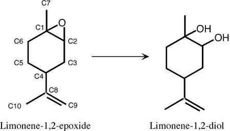
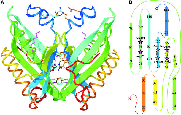
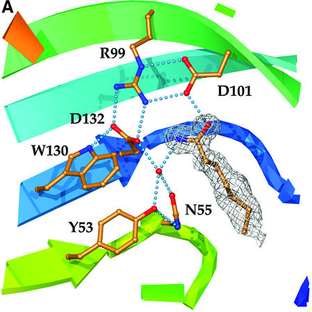
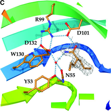
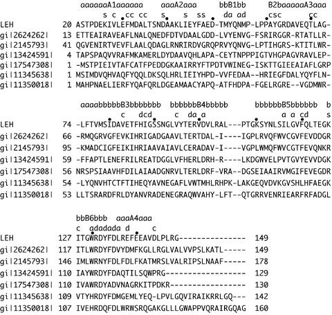
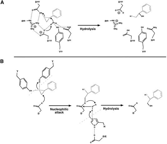
Epoxide hydrolases are essential for the processing of epoxide-containing compounds in detoxification or metabolism. The classic epoxide hydrolases have an alpha/beta hydrolase fold and act via a two-step reaction mechanism including an enzyme-substrate intermediate. We report here the structure of the limonene-1,2-epoxide hydrolase from Rhodococcus erythropolis, solved using single-wavelength anomalous dispersion from a selenomethionine-substituted protein and refined at 1.2 A resolution. This enzyme represents a completely different structure and a novel one-step mechanism. The fold features a highly curved six-stranded mixed beta-sheet, with four alpha-helices packed onto it to create a deep pocket. Although most residues lining this pocket are hydrophobic, a cluster of polar groups, including an Asp-Arg-Asp triad, interact at its deepest point. Site-directed mutagenesis supports the conclusion that this is the active site. Further, a 1.7 A resolution structure shows the inhibitor valpromide bound at this position, with its polar atoms interacting directly with the residues of the triad. We suggest that several bacterial proteins of currently unknown function will share this structure and, in some cases, catalytic properties.


Figures

None
Fig. 1. The reaction of LEH with its natural substrate. Carbon atoms are numbered.
None
Fig. 2. Structure of LEH. (A) Ribbon drawing showing the dimer, with each subunit coloured going through the rainbow from red at the N-terminus to blue at the C-terminus. Some of the residues contributing to the dimer interface, as described in the text, are shown as ball-and-stick representations. The endogenous ligand is also shown (magenta) in both subunits. (B) Topology diagram of the LEH subunit, using the same rainbow scheme. Residues included in each secondary structural element are numbered; helix α2 is irregular. Active-site residues are indicated by magenta stars.
None
Fig. 3. The active site. (A) Catalytic residues, showing their relationship to each other and supporting side-chains, as well as to the water molecule and endogenous ligand found in the LEH active site. Hydrogen-bonding interactions are shown by dotted lines. Colouring of the ribbon portions follows the rainbow scheme defined in Figure 2. The electron density of the endogenous ligand (modelled as heptanamide) in the final 2Fo – Fco map is contoured at a level of 1σ. (B) Hydrogen-bonding interactions between active-site groups and the endogenous ligand. The figure shows Asp132 acting as the base and Asp101 as the acid. Where the donor–acceptor relationship is not clear from the available data, hydrogen bonds are indicated by double-headed arrows. (C) Complex with valpromide. The electron density of the final 2Fo – Fco map is contoured at a level of 1σ.
None
Fig. 4. Sequence alignments of LEH with proteins of unknown function. Sequence alignment was performed using hidden Markov models (Karplus et al., 1998), with some small manual adjustments using the LEH structure as a guide. Every tenth residue in the LEH sequence is marked. Residues preceding α1 are poorly conserved or missing in the other proteins, and so the alignments shown omit those segments. The roles that conserved residues play in LEH are indicated using the following code: a, active site; c, core or other structure; d, dimer; s, surface. Members of the first group of proteins, which appear to have both structural and functional relationships to LEH, are gi|2145793| (hypothetical protein B2235_F3_140 from Mycobacterium leprae), gi|2624262| (hypothetical protein Rv2740 from Mycobacterium tuberculosis), gi:13424591| (hypothetical protein from Caulobacter crescentus) and gi:17547308 (conserved hypothetical protein from the plant pathogen Ralstonia solanacearum). Members of the second group, which are presumed to have a similar structure, but distinct function, are gi|11345638| (hypothetical protein VC1118 from Vibrio cholerae) and gi|11350018| (hypothetical protein PA3856 from Pseudomonas aeruginosa). A few regions may contain small shift errors, which will require new sequence/structural data to locate and correct.
None
Fig. 5. Mechanisms of epoxide hydrolases. (A) The mechanism of LEH indicated by the experimental results, using styrene oxide as an example. The catalytic water molecule is held in place and activated by hydrogen bonding to residues Asp132, Asn55 and Tyr53. The activated water molecule forces epoxide ring opening by nucleophilic attack at one of the ring carbons. At the same time, Asp101 activates the epoxide ring by donation of a proton to the epoxide oxygen (acid catalysis). Thus the formation of the diol from the epoxide proceeds in a single step by a push–pull mechanism. After this step, Asp132 should be in the protonated state and Asp101 should be charged, which can be rapidly reversed with the aid of Arg99 as a proton shuttle. The hydrogen bond donor/acceptor atoms for Tyr53 and Asp55 cannot be proved using current information, and only one of the two possibilities is drawn. (B) Reaction mechanism of α/β fold EHs. In these enzymes, two tyrosines position the epoxide oxygen by hydrogen bonding and activate the epoxide for nucleophilic attack by an aspartic acid residue. This first step leads to the formation of an enzyme–substrate ester intermediate. Subsequent hydrolysis of the intermediate is achieved by a water molecule activated by a His-Asp/Glu charge relay system. The hydrolysis leads to product formation and reconstitution of the active enzyme. The tyrosines and charge relay system are only shown in the present scheme where they contribute to the mechanism.

References

    1. Altschul S.F., Madden,T.L., Schaffer,A.A., Zhang,J., Zhang,Z., Miller,W. and Lipman,D.J. (1997) Gapped BLAST and PSI-BLAST: a new generation of protein database search programs. Nucleic Acids Res., 25, 3389–3402. - PMC - PubMed
    1. Arand M., Hemmer,H., Dürk,H., Baratti,J., Archelas,A., Furstoss,R. and Oesch,F. (1999) Cloning and molecular characterization of a soluble epoxide hydrolase from Aspergillus niger that is related to mammalian microsomal epoxide hydrolase. Biochem. J., 344, 273–280. - PMC - PubMed
    1. Archelas A. and Furstoss,R. (2001) Synthetic applications of epoxide hydrolases. Curr. Opin. Chem. Biol., 5, 112–119. - PubMed
    1. Argiriadi M.A., Morisseau,C., Hammock,B.D. and Christianson,D.W. (1999) Detoxification of environmental mutagens and carcinogens: structure, mechanism and evolution of liver epoxide hydrolase. Proc. Natl Acad. Sci. USA, 96, 10637–10642. - PMC - PubMed
    1. Argiriadi M.A., Morisseau,C., Goodrow,M.H., Dowdy,D.L., Hammock,B.D. and Christianson,D.W. (2000) Binding of alkylurea inhibitors to epoxide hydrolase implicates active site tyrosines in substrate activation. J. Biol. Chem., 275, 15265–15270. - PubMed

Publication types